Book Now

The Hidden Truth About Calcaneal Spur Surgery: What Your Doctor May Not Tell You

calcaneal spur surgery

A calcaneal spur, often referred to as a heel spur, is a bony growth that develops on the heel bone (calcaneus). This condition is commonly associated with plantar fasciitis, which involves inflammation of the plantar fascia ligament, leading to significant heel pain. While many individuals manage heel pain through conservative treatments, some may require calcaneal spur surgery to alleviate persistent discomfort and restore mobility. This blog aims to provide a better understanding of why the removal of the heel spur is rarely needed and can be considered a fallacy. I will contrast the old-school ideas with the more recent concepts based on science.

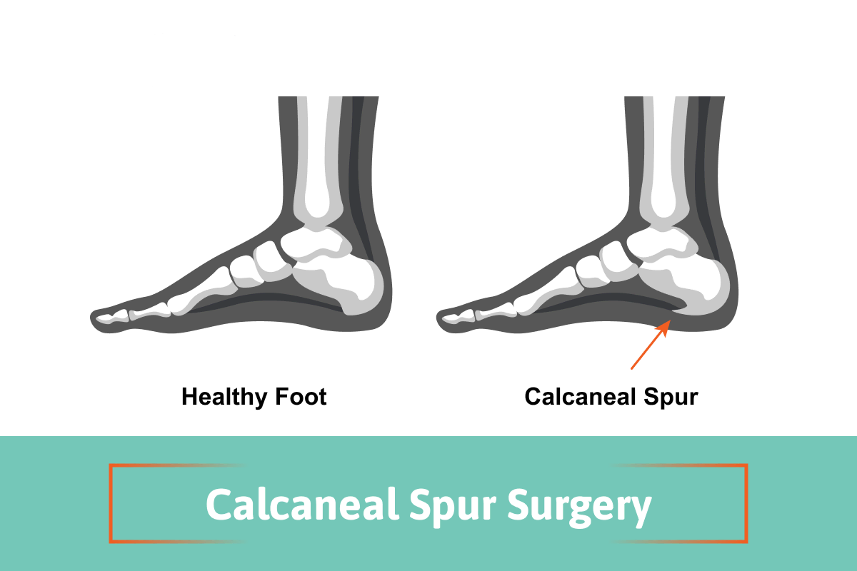
What is a Calcaneal Spur?

A calcaneal bone spur is a bony growth that forms on the underside of the heel bone, typically at the site where the plantar fascia attaches to the calcaneus. Most believe that the point of the fascia from the heel bone at this location may stimulate new bone to form, thus causing the spur formation. In the 1930s, Dr. Spitz was the first doctor to suggest that the actual heel spur is not causing the pain, but rather the fascia that is pulling from the calcaneus.

This condition can arise due to repetitive stress and strain on the foot, often resulting from activities such as running, walking, or standing for long periods. The bone spur development can occur alongside plantar fasciitis, leading to significant heel pain and discomfort. Symptoms may include sharp pain in the heel, especially in the morning or after prolonged periods of sitting. While many individuals experience relief through non-surgical interventions, some may require surgery when conservative treatments fail to provide adequate relief.

Causes of Calcaneal Spurs

calcaneal spur surgery causes infographic

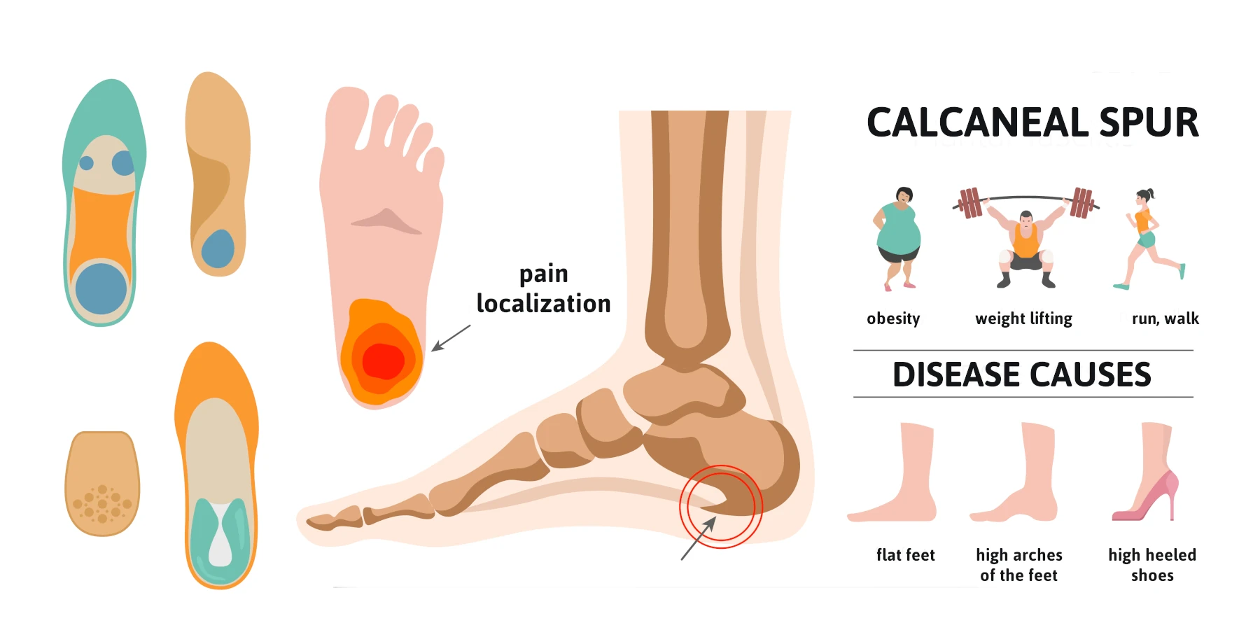
Calcaneal spurs commonly develop due to a combination of factors, including:

  • Repetitive Stress: Activities that involve excessive strain on the feet, such as running, dancing, or standing for extended periods, can lead to the formation of a heel spur.
  • Plantar Fasciitis vs Plantar Fasciosis: the old school concept is that the fascia is inflamed. This has been disproven by one study performed by Dr. Harvey Lamont. When he performed surgery to release the plantar fascia from the heel bone, specimens were sent to a lab. Out of 50 specimens, 49 showed fascial degeneration while only one showed inflammation. This supports that it is not an inflammatory condition.
  • Chronic pulling of the plantar fascia can contribute to the development of a calcaneal spur, as the body attempts to stabilize the area by forming new bone.
  • Foot Structure: Abnormalities in foot structure, such as flat feet or high arches, can increase the risk of developing heel spurs.
  • Age and Weight: Excess body weight can contribute to higher stress levels on the feet.

Symptoms of Calcaneal Spurs

Individuals with a calcaneal spur often experience a range of symptoms, including:

  • Heel Pain: The most common symptom is a sharp, stabbing pain in the heel, especially during the first steps in the morning or after prolonged sitting.
  • Swelling and Inflammation: The area around the heel may become swollen and tender to the touch due to inflammation.
  • Pain with Activity: Activities like walking, running, or standing for long periods can exacerbate heel pain.
  • Difficulty in Movement: In severe cases, individuals may find it challenging to walk or engage in physical activities due to discomfort. Understanding these symptoms is crucial for individuals to seek appropriate medical evaluation and treatment.

Diagnosis of Calcaneal Spurs

Diagnosing a calcaneal spur typically involves a thorough evaluation by a healthcare professional. The diagnostic process may include:

  • Medical History: The doctor will inquire about the patient’s symptoms, pain levels, and any previous foot injuries or conditions.
  • Physical Examination: A physical examination of the foot will help assess tenderness, swelling, and range of motion.
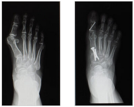
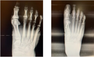
  • Imaging Tests: X-rays are often used to confirm the presence of a calcaneal spur and assess its size and location. In some cases, MRI or ultrasound may be utilized to evaluate soft tissue involvement, such as the plantar fascia and the Achilles tendon. An accurate diagnosis is essential for determining the most appropriate treatment options, whether conservative or surgical.

Non-Surgical Treatment Options

Before considering surgery, healthcare providers typically recommend conservative treatment options for calcaneal spurs, including:

  • Rest and Activity Modification: Reducing high-impact activities and allowing the foot to heal can alleviate symptoms.
  • Physical Therapy: A physical therapist can design a personalized program that includes stretching and strengthening exercises for the foot and ankle, improving flexibility and reducing pain.
  • Orthotic Devices: Custom orthotics or shoe inserts can provide additional support and cushioning to the foot, helping to redistribute pressure away from the heel.
  • Pain Medication: Over-the-counter medications, such as ibuprofen or acetaminophen, can help manage pain and inflammation.
  • Cortisone injection vs Regenerative medicine: Corticosteroid injections may be helpful but rarely provide permanent relief and this is because the fascia is not inflamed, but rather degenerating based upon studies. Therefore, the option of regenerative medicine provides a very success rate to reverse the symptoms on a long-term basis as this treatment helps repair the fascia that is degenerating.

When is Surgery Necessary?

Surgery for calcaneal spurs is generally considered when conservative treatments fail to alleviate pain and improve mobility after several months. Indications for surgery may include:

  • Persistent Pain: Severe, ongoing heel pain that significantly affects daily activities and quality of life.
  • Diagnosis Confirmation: Confirmation of a calcaneal spur through imaging tests and persistent symptoms despite conservative management.
  • Functional Limitations: Difficulty participating in work, recreational activities, or exercise due to heel pain. Surgical intervention aims to remove the spur and address any accompanying issues, such as plantar fasciitis, to restore function and reduce pain.
  • Regenerative Medicine: The option of regenerative medicine has greatly reduced the need for surgery. (see testimonial videos)

The Surgical Procedures for a Calcaneal Spur

Contrasting heel spur surgery to endoscopic plantar surgery. Calcaneal heel spur surgery usually involves incision on the side of the heel and the spur is removed along with the attachment of the fascia to the heel bone at the spur location. This is a procedure that surgeons have not performed since 1990. Because the heel spur is not thought to be the cause of your pain, using a small incision to place a scope across the bottom of the heel allows the surgeon to cut a portion of the fascia so that it may stretch and elongate and reduce its pull on the heel bone. This technique has been adopted by majority of the Podiatric Foot and Ankle Surgeons, and because of its high success rates, it supports that fact that the heel spur is not the cause of your pain.

Recovery from heel spur surgery

This type of surgery usually involves a larger incision and more trauma and longer recovery with immobilization in a cast, and sometimes non-weight bearing.

Recovery from endoscopic heel surgery

This type of surgery uses a small incision, and you can bear weight immediately and may return to wearing shoes in 3-4 weeks.

Watch Patient Testimonial Videos


Post-surgery, patients are typically advised to avoid weight-bearing activities for a specified period to allow for proper healing. Physical therapy may be recommended to facilitate full recovery and regain strength and range of motion.

Recovery and Rehabilitation for Calcaneal Spur Surgery

The recovery time for calcaneal spur surgery varies among individuals, with most patients experiencing significant improvement within a few weeks. A typical recovery process includes:

  • Pain Management: Pain may be managed with prescribed medications and the application of ice to reduce swelling.
  • Physical Therapy: Engaging in physical therapy is crucial for restoring strength, flexibility, and balance. Therapists may focus on exercises to improve range of motion and stability, particularly around the Achilles tendon and plantar fascia.
  • Gradual Return to Activity: Patients are typically encouraged to gradually return to normal activities as healing progresses, following their surgeon’s recommendations.
  • Long-Term Management: Maintaining a healthy weight, wearing appropriate footwear, and incorporating regular stretching and strengthening exercises can help prevent future heel issues.

Conclusion

Calcaneal spur surgery can provide significant relief for individuals suffering from persistent heel pain due to calcaneal spurs. While many patients find success with conservative treatments, surgery remains an effective option for those who do not respond to traditional methods. Understanding the causes, symptoms, and treatment options for calcaneal spurs is essential for making informed decisions about care. If you suspect you have a calcaneal spur or are experiencing persistent heel pain, consult with a healthcare professional to explore the best treatment options for your needs.

Bunion Surgery Recovery: A Complete Week-by-Week Breakdown

Bunion Surgery week by week

Bunion surgery, or a bunionectomy, can be a life-changing procedure for individuals suffering from painful, misaligned toes. While the surgery itself is relatively common, many patients are unsure about what to expect during recovery. Healing from bunion surgery takes time, and understanding the recovery process week by week can help you prepare both physically and mentally. Here’s what you can expect after bunion correction surgery.

Bunion Surgery Recovery Basics

Before you read the week-by-week recovery that you would expect from bunion surgery, some basic principle bunions need to be understood. With very few exceptions when surgery is performed, the first metatarsal bone needs to be shifted towards the second metatarsal bone to correct the increased angle between the two bones.

This increased angle is what causes the bunion protrusion. This requires that the bone heals in a more correct position. The surgical technique to shift the bone can take place at the level of the metatarsal head which is close to the big toe joint or back at the first metatarsal base which is towards the mid-portion of your foot. Approximately 90% of surgeries we perform are in the area of the metatarsal head and most patients can return to regular shoes between 3 and 4 weeks after surgery. For more severe bunions, they may have to be off weight bearing for 5-6 weeks. This is important to understand because when the bunion starts to hurt, it may be advantageous to get it corrected sooner than later.

The majority of these surgeries take place in an out-patient setting and no general anesthesia is needed. The surgery can be done in a minimally invasive approach and may take approximately an hour and local anesthetic, and IV sedation is given during the surgery. The recurrence rate after the surgery is extremely low, and the patients are able to return to their normal daily activities after weeks post-surgery.

(Watch Bunion Surgery Patient Testimonial Videos)


Week 1: Immediate Post-Surgery Care

The first week is all about rest and protecting your affected foot. You’ll likely experience swelling, pain, and discomfort, which is managed with prescribed pain medication and keeping your foot elevated.

Key Instructions:

  • Keep your foot elevated as much as possible (above heart level) to reduce swelling.
  • Use ice packs periodically to manage pain and inflammation.
  • Avoid putting weight on the affected foot; use crutches or a walker as advised.
  • Keep the surgical dressing dry and intact to prevent infection.
  • Follow up with your surgeon for post-operative instructions.
  • Be aware of deep vein thrombosis risks and watch for unusual swelling or pain.

Week 2: Early Healing and Mobility Adjustments

By the second week after surgery, some pain and swelling may persist, but you should notice gradual improvement. Your stitches may be removed during this period, depending on the types of bunion surgery performed.

Key Instructions:

  • Continue keeping your foot elevated when sitting or lying down.
  • Start moving your big toe slightly if recommended by your doctor to maintain range of motion.
  • Some patients may transition to wearing surgical shoes instead of using crutches.
  • Watch for signs of infection, such as excessive redness, warmth, or drainage.

Week 3-4: Reduced Swelling and Increased Activity

At this stage, you may start feeling more mobile, but your foot and ankle are still healing

Key Instructions:

  • Swelling may still be present, but it should be decreasing.
  • Depending on the procedure, you may begin some weight bearing on the affected foot.
  • You might start gentle range-of-motion exercises to prevent stiffness.
  • Avoid prolonged walking or standing.
  • Keep wearing your surgical shoes as directed.

Week 5-6: Transitioning to Normal Activities

This is when many patients begin to feel significantly better and may even start wearing regular shoes.

Key Instructions:

  • You may transition to a more supportive shoe with a wide toe box.
  • Swelling is still common, especially by the end of the day.
  • You may be cleared for physical therapy to improve strength and mobility.
  • Avoid high-impact activities like running or jumping.

Week 7-8: Resuming Light Exercise and Daily Routines

At this point, many patients find they can walk more comfortably, though swelling may persist.

Key Instructions:

  • Walking distances can gradually increase.
  • Light stretching and strengthening exercises may be introduced.
  • Swelling may still occur, especially after long periods of standing.
  • You might be able to return to work if it doesn’t require extensive foot use.

Week 9-12: Near Full Recovery

By the end of three months after surgery, most patients can return to normal activities with minimal discomfort.

Key Instructions:

  • You should be able to walk normally in comfortable footwear.
  • Continue exercises to maintain mobility and strength.
  • Some residual swelling may persist but should be much less frequent.
  • High-impact activities may be gradually reintroduced with your doctor’s approval.

Month 4-6: Long-Term Healing

Though you may feel fully healed, complete recovery from bunion surgery can take up to a year.

Key Instructions:

  • Continue to wear supportive footwear to prevent recurrence.
  • Regular stretching and strengthening exercises help maintain flexibility.
  • Swelling should be minimal or nonexistent.
  • High-impact sports and activities can typically resume.

Final Thoughts

It is important to understand that most patients we see for bunions come to us because they are painful, and by that time, the conservative treatment options are less likely to be recommended. If the bunion deformity is dealt with earlier before there is pain, orthotic devices (custom-made arch supports) can help with the discomfort and have some effect on slowing the progression of the condition. Unfortunately, most people allow their pain to be their guide and don’t come in earlier where orthotic treatments could still be useful. Orthotics are also useful after the surgery especially in younger people to alter the biomechanics of the foot that created the bunion to begin with.

Recovery from bunion surgery varies from person to person, but patience is key. Following your doctor’s instructions, keeping up with physical therapy, and taking care of your feet will ensure the best possible outcome. If you experience unusual pain, swelling, or difficulty moving your foot, consult your surgeon promptly.

With the right approach, you’ll be back on your feet—pain-free—before you know it!


The Perfect Read If You’re Considering Bunion Surgery Lapiplasty!

bunion surgery lapiplasty

Bunions are a deformity of the big toe joint that can make it difficult and painful to wear shoes. This affliction may be addressed by doing a bunion surgery lapiplasty correction when conservative options fail. If you’re noticing a bump on the inside of your big toe joint that is bothering you when you wear fashionable shoes – this blog is for you.

What is a Bunion?

A bunion deformity is caused by a misalignment of the bones in the foot. The first toe will angulate towards the second toe and the first metatarsal will angulate away from the second metatarsal.

Normally, the first and second metatarsal bones should be close to parallel to each other. However, with a bunion deformity, the angle between the two is increased. This will give the appearance of a large bump on the inside of your big toe joint. The actual bump can be from extra bone on the inside of what is referred to as the metatarsal head but more accurately the primary cause of the prominence is the angle between the metatarsal bones.

What is the goal of surgery?

The goal of surgery is to correct the angle between the first and second metatarsal bones. This correction brings the angle back to a normal anatomical alignment. We do this in such a way that there is no recurrence.

The History of Bunion Correction Procedures

Decades ago, many patients who had bunion surgery had the bump removed. They would use the soft tissue around the great toe joint to realign the toe. These procedures made for an easy recovery but resulted in minimal reduction of deformity and a very high recurrence rate.

bunions-infographic

Metatarsal Osteotomies

Next, foot and ankle surgeons started to do procedures with osteotomies on the first metatarsal bone to correct the deformity and remove the bump. An osteotomy means that the bone is cut and realigned or shifted to correct the angulation. There were two types of osteotomies; those performed at the metatarsal head (Austin or chevron) in order for the metatarsal head to be shifted. Otherwise, the osteotomies are performed at the base of the metatarsal bone. Those done at the base were for more severe bunions. These types of procedures involved significantly more lay up time. Whether the correction of the deformity is performed at the metatarsal head or the metatarsal base, they are considered traditional bunion surgery.

Next, surgeons determined that maybe the best surgical option was to fuse the first metatarsal cuneiform joint because this is considered an unstable joint. This is called the Lapidus procedure. It corrects the misalignment with a joint fusion. This can work very well but it includes a long lay time. Patients are not allowed to put weight on their foot for at least six weeks non weight bearing. There was also a risk of nonunion or delayed union of bone at the fusion site.

Bunion Surgery Lapiplasty

The bunion surgery lapiplasty procedure is a refinement of the Lapidus procedure. It is promoted as correcting the deformity in three different planes. The procedure also secures the unstable foundation which is believed to be the first metatarsal cuneiform joint. It also allows for early weight bearing. However, it is recommended that you be in a Cam walker so the bones are fully healed for at least six weeks. It is also promoted as having minimal recurrence rate. There are also claims that doing osteotomies of the metatarsal head have very high recurrence rates. Lapiplasty is promoted as having advanced fixation technology to fuse the joint. This technique uses titanium plating technology and these anatomically shaped titanium plates remain in the foot after surgery. Lastly, specialized instrumentation to make the results more precise and predictable are used.

Dr. Anderson’s Opinion of Bunion Surgery Lapiplasty

First, understand that I’ve been curious about this procedure but yet have not performed this procedure.

Here are some notes:

Recurrence Rates: In the marketing material for the Lapiplasty, they suggest a very high recurrence rate for patients that have an osteotomy of the metatarsal head. As a surgeon that has been doing bunion surgeries for 4 decades, I differ with this. Very seldom do we see recurrences. If surgeons were more like me, we would not have continued with these techniques.

They suggest that the correction is more precise because the bunion is being corrected in three planes and not two. In regards to this, yes, I can definitely agree with this; however, one would then assume that if the correction is not done in this manner, less correction was made, and recurrence rates were high – and on this I disagree.

With the Lapiplasty procedure, it is suggested that you will have less layup time. You’ll be able to bear weight within days and will ambulate in a Cam walker for 6 weeks. I’m not sure where the narrative comes from but most patients’ assumption of how long they are laid up is significantly more than the reality. The current way that we do most of our bunion corrections is a few days of non-weight bearing, then walking in a surgical boot or surgery shoe for up to 3-4 weeks. Patients can return to regular shoes in 3-4 weeks. Therefore the conventional approach to bunion surgery using a metatarsal head osteotomy is significantly less than the bunion surgery lapiplasty.

X-Rays of bunion surgery lapiplasty

X-Rays of Anderson Podiatry Center Technique

  • Majority of patients opt for our procedure due to successful outcomes
  • No pins left in the foot
  • Minimal chance of recurrence
  • You are back to wearing shoes faster

Hardware Complications: Currently we use small pins to fixate your bone with our bunion corrections. The advantage is that they are removed just weeks after, through a very small incision. Therefore, you don’t need to worry about potential pain from bone screws or plates that remain in your foot.

Marketing for Doctors: in the competitive medical world doctors are sometimes looking for an approach that sets them apart or allows them to market themselves. I have no problem with this but if you’re considering this procedure, please understand that there is motivation to do this procedure because the company that makes the instrument is marketing the doctors. You do the procedure and we will market your practice for you to generate more patients. Please consider this.

My Conclusion

I think the Lapiplasty bunion correction is a welcome addition for how foot and ankle surgeons correct a bunion. I don’t think it should be a one size fits all to be done on all bunions. I also think that it’s misleading to think that this procedure will lay you up less, as with many current approaches you may be back in shoes in 3-4 weeks, not 6 weeks. Many patients don’t know enough about bunion surgery to understand that indeed their bunion deformity may not require the procedures that require longer lay ups. In fact, the majority do not. I can agree that the technique used may lead to more consistent results, that’s assuming the conventional approaches have a lot of variation in outcomes. To summarize, you need to question the recurrence rates that they claim with conventional surgeries, and if it is worth the longer lay up times. If you are considering bunion correction please don’t hesitate to contact us for an initial bunion foot evaluation.

We have local foot clinics in the Fort Collins and Broomfield area with certified podiatrist who are experienced in bunion corrections. Watch out patient testimonial below to hear about the success, Phyllis is just one of many we have treated over the years.

For a full list of videos view our Bunion You Tube playlist.

Watch Videos

What You Need to Know About Foot Surgery for Bunions and Three Ways They are Corrected!

foot-surgery-for-bunions

Bunions can be a painful foot condition for many. They can become painful and make finding a comfortable pair of shoes to wear very difficult. Foot surgery for bunions should be not only be considered for the asthetical value, but the overall bony health and balance of the body.

What is a Bunion & What Causes a Bunion?

There is a primary problem that creates a bunion, which starts as the big toe begins to anglalate towards the smaller toes. The metatarsal bone at the base of the digit moves away from the rest of the foot causing an increased angle. This increased angle is between the first and second metatarsal bones. The results are a hard bony prominence at the base of the big toe. The medical term for this condition is Hallux Valgus.

The primary cause is thought that poor foot mechanics may be the main cause. Too much weight may be placed beneath the great toe joint, and it changes the pull of the tendon around the great toe which starts the process. Also, wearing shoes such as high heels that are pointed or too tight may also be a cause.

Non Surgical Treatments

When it comes to bunions, there are multiple non-surgical options to tryout:

  1. Change in Shoe Gear – Try to avoid shoes that are too tight and purchase shoes with a larger toe box
  2. Padding – Placing more cushion on the area may also provide temporary relief and the use of anti-inflammatory drugs
  3. Orthotics – Custom made shoe inserts called orthotics may also be useful

Surgical Treatment Option for Bunions

For many, the surgical correction becomes the treatment of choice. When it’s difficult to wear the majority of shoes this may be the appropriate treatment.

We can discuss three basic approaches to correcting the bunion. To begin with the problem lies in the angulation of the first metatarsal away from the second metatarsal bone. This is what causes most of the bony prominences that rub against the shoes. This angle needs to be reduced so the two bones are more parallel. By accomplishing this angle correction, it is easier to straighten the big toe. Many years ago, bunion surgery had a bad reputation because doctors would simply remove the bone bump and not address the angulation of the bone. Thus, for many, this only offered part-time relief.

How is the angle corrected? It can be done in three general ways:

  • By shifting the bone close the great toe joint
  • By shifting the bone by the base of the metatarsal bone closer to the instep
  • By removing a portion of the great toe joint

It is also important to note that what procedure the foot and ankle surgeon choses is based upon the severity of the bunion deformities and the patient’s age.

Three Types of Foot Surgery for Bunions Performed

  1. There is the option of shifting the bone by the great toe joint. The vast majority of toe time, this procedure is performed at Anderson Podiatry Center. The technique is used for more mild or moderate bunions. The recovery from this is usually wearing a surgical shoe or Cam walker for 3-4 weeks and just a few days of non-weight being in the beginning.
  2. The option of shifting the bone at the base closer to the ankle usually requires more lay-up time of weeks after surgery, usually 4-6 of off- weight bearing. This procedure is reserved for more severe deformities and may only comprise ten percent of the bunion corrections that we do.
  3. This final option is to remove a portion of the great toe joint. This can work very well in the senior population over 65 years of age. There is very little risk and has been performed successfully in patients in their 80’s. For all these surgical options, the affected foot will need to be elevated after surgery for several days and weight bearing can resume within days or weeks depending on what surgical approach is performed. Physical therapy may also be recommended during the surgical recovery. When you can return to normal activity level will also vary.

Below, I have a few notes in regards to bunion surgery and occurrence.

Surgical Setting: These surgeries are performed on an outpatient basis using local anesthesia and IV Sedation. Depending on the circumstance, both feet may be done at once.

Recurrence Rates: The possibility of recurrence of the bunion is very minimal especially if the foot and ankle surgeon is shifting the first metatarsal bone, and not simply removing the bunion bump.

Why Early Treatment May be Advantageous

Foot Surgery for bunions should be carefully considered and should not be done for cosmetic reasons. Once there is pain and shoes are becoming a challenge, surgery before the bunion becomes more severe may be best. Why? Simply because of lay-up time. This is something few patients are aware of. Many assume it may be weeks of not bearing weight on the foot. This is not the case for many patients. The majority of corrections we perform allow you to place weight on the foot within a few days.

Call us today at our Fort Collins location (970) 484-4620, Broomfield location (720) 259-5053 or use our online scheduling system to book your appointment.

We can find a solution for you!

Bunion pain relief ! Karen is back to wearing the shoes she loves. She shares her experience.

Bunion Surgery Success! Tammy Shares Her Story

Susan’s shows off her shoes she can now wear . Bunions fixed !

Bunion surgery on both feet! Kari shares her honest story of what it was like for her.
Bunion Surgery – Kari