Book Now

Diabetic Nerve Pain in Feet: New Hope Through Advanced Treatments

Nerve Pain In Feet

Living with diabetes brings many challenges, and diabetic nerve pain in feet ranks among the most debilitating complications. This condition, called diabetic neuropathy, affects millions of people with diabetes worldwide. The good news is that recent advances in treatment offer new hope for managing this painful condition.

Understanding Diabetic Neuropathy

Diabetic neuropathy occurs when high blood sugar levels damage nerves throughout your body over the long term. The most common type is diabetic peripheral neuropathy, which primarily affects nerves in your feet and legs. This nerve damage can severely impact your ability to feel pain, temperature, and touch.

The condition affects nerves that control various body functions. When blood vessels that supply these nerves become damaged, it leads to the characteristic symptoms many patients experience. Managing your blood sugar levels is crucial, as elevated glucose damages both blood vessels and nerve fibers over time.

Types of Diabetic Neuropathy

There are several types of diabetic neuropathy, and symptoms depend on the type affecting you:

Peripheral Neuropathy: The most common type, causing numbness, burning sensations, and sharp pains in feet and legs. Many patients report symptoms become worse at night.

Autonomic Neuropathy: This type affects nerves that control internal organs, including blood pressure heart regulation, digestive system, and sex organs. It can impact bladder control and sexual function.

Proximal Neuropathy: Affects nerves in the hips, thighs, and buttocks, causing sudden weakness and pain.

Focal Neuropathy: Targets specific nerves, creating localized weakness or pain in particular areas.

Understanding which type affects you helps doctors create the most effective treatment plan for your specific situation.

Why Feet Are Most Vulnerable

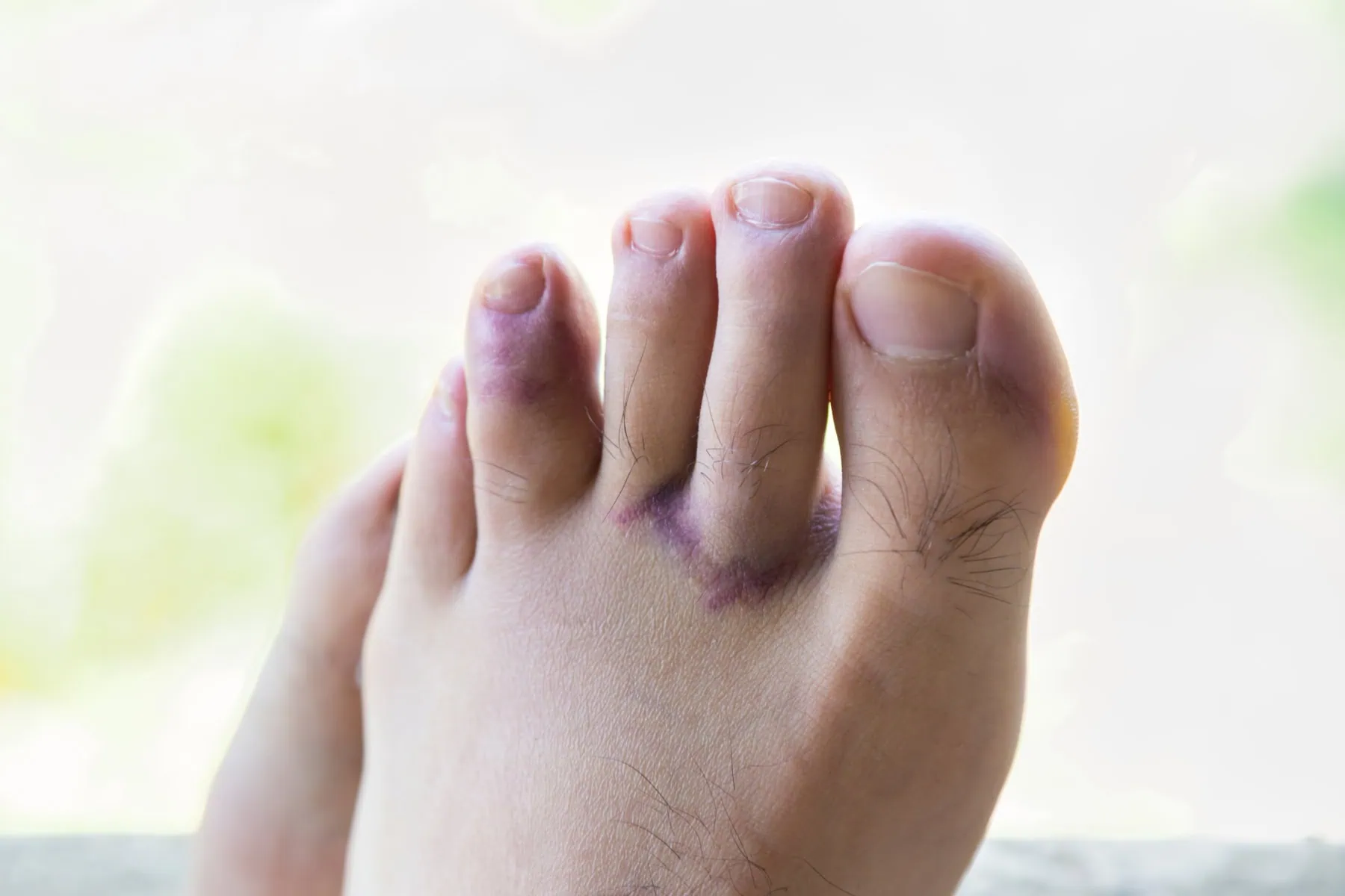
Your feet bear the brunt of diabetic neuropathy for several reasons. The longest nerves in your body extend to your feet, making them most susceptible to damage from high blood sugar. Additionally, blood vessels supplying these distant nerves often sustain damage first, reducing oxygen and nutrient delivery to nerve tissues.

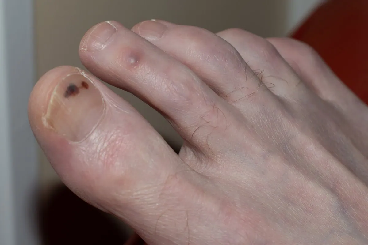
This nerve damage impairs your ability to feel pain properly, which normally serves as a protective warning system. Without this protection, minor injuries can go unnoticed and develop into serious complications.

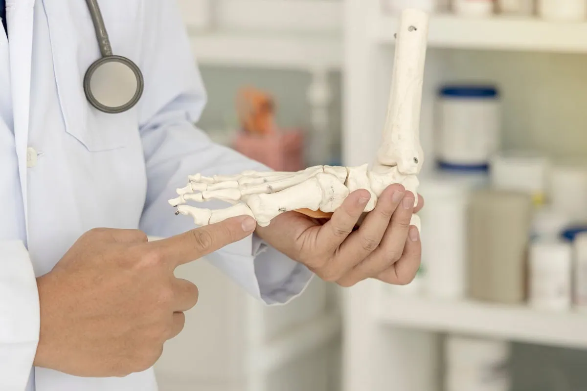
Breakthrough Research: Dr. James Anderson’s Nerve Decompression

Recent groundbreaking research by Dr. James Anderson, DPM, has revolutionized treatment approaches for diabetic nerve pain in feet. His work focuses on nerve entrapment, where damaged nerves become compressed in anatomical tunnels throughout the lower legs and feet.

Dr. Anderson’s research demonstrates that nerve decompression surgery can significantly improve symptoms by relieving pressure on compromised nerves. This surgical technique targets specific compression points, improving blood flow and reducing nerve irritation.

Benefits of Nerve Decompression Surgery

Patients who undergo Dr. Anderson’s nerve decompression procedures often experience:

  • Restored sensation in feet and toes
  • Significant reduction in burning and stabbing pains
  • Lower risk of foot ulcers and potential amputations
  • Improved balance and walking stability
  • Potential restoration of some lost nerve function

Early intervention produces the best outcomes, emphasizing the importance of seeking treatment before irreversible damage occurs.

Essential Foot Care for People with Diabetic Nerve Pain In Feet

Proper foot care remains fundamental for anyone managing diabetic peripheral neuropathy. Daily inspection helps identify problems early, before they become serious complications.

Daily Foot Care Routine:

  • Examine feet thoroughly for cuts, blisters, or color changes
  • Keep feet clean and completely dry
  • Wear properly fitted, supportive footwear
  • Never walk barefoot, even indoors
  • Moisturize feet but avoid areas between toes

These simple steps can prevent minor issues from becoming major complications that threaten your mobility and independence.

Comprehensive Treatment Approaches For Diabetic Nerve Pain In Your Feet

Managing diabetic nerve pain in feet requires a multi-faceted approach combining various treatment options:

Blood Sugar Management: Maintaining stable blood sugar levels remains the cornerstone of preventing further nerve damage. Work closely with your healthcare team to optimize your diabetes management plan.

Medication Options: Several medications can help manage neuropathy pain, including specific pain relievers, certain antidepressants, and anti-seizure medications that affect nerve pain signals.

Physical Therapy: Targeted exercises improve strength, balance, and mobility while helping maintain nerve function in affected areas.

Topical Treatments: Creams containing capsaicin or lidocaine patches can provide localized pain relief for some patients.

Nutritional Support: Certain vitamins and supplements may support nerve health, though you should discuss these options with your physician before starting any new supplements.

Managing Related Health Concerns

Diabetic neuropathy often affects more than just your feet. Autonomic neuropathy can impact blood pressure heart function, digestive processes, and sex organs. Working with your healthcare team to address these interconnected issues improves your overall quality of life.

Regular monitoring of blood pressure, heart health, and other organ functions helps detect and treat complications early. This comprehensive approach ensures you receive the most effective care for all aspects of your condition.

Hope for the Future

Thanks to pioneering research from specialists like Dr. James Anderson, treatment options for diabetic nerve pain in feet continue expanding. Nerve decompression surgery represents just one exciting advancement offering hope for symptom improvement and potential nerve function restoration.

The key to success lies in early intervention and comprehensive care. If you experience symptoms of diabetic neuropathy, don’t wait for them to worsen. Speak with your healthcare provider about all available treatment options, including advanced surgical techniques that might be appropriate for your situation.

With proper foot care, diligent blood sugar management, and access to cutting-edge treatments, living well with diabetic neuropathy is increasingly possible. Take control of your condition today by exploring all available options and working with specialists who understand the latest advances in neuropathy care. View more informational and testimonial videos on our YouTube Channel.

Watch Our Informational Videos On Nerve Pain From Diabetic Neuropathy

Schedule Your Appointment To Treat Diabetic Nerve Pain In Feet

Call us today at our Fort Collins location (970) 484-4620, Broomfield location (720) 259-5053 or use our online scheduling system to book your appointment.

Learn the Causes and Treatments for Thick Yellow Toenails

Thick Yellow Toenails

Thick yellow toenails can be more than just a cosmetic issue — they often signal an underlying problem that requires attention. Whether it’s caused by a fungal nail infection or another condition, yellow toenails should never be ignored. In this blog, we’ll cover the common causes, prevention strategies, treatment options, and home remedies, including how products like tea tree oil can play a role. If you or someone you know struggles with thick, discolored toenails, this guide is for you.

What Causes Thick Yellow Toenails?

Several factors can lead to the development of thick, yellow toenails, but the most common cause is toenail fungus. Fungi thrive in warm, moist environments, making feet the perfect breeding ground, especially when you wear shoes for extended periods or walk barefoot in public places like locker rooms. Other causes include:

  • Yellow nail syndrome, a rare disorder associated with respiratory problems and swelling.
  • Psoriasis or other skin conditions affecting the nail bed.
  • Aging, where nails naturally thicken and discolor over time.
  • Compromised immune system function, making it easier for infections to take hold.

Regardless of the cause, noticing an infected toenail early can make treating toenail conditions much easier.

Recognizing Symptoms of Nail Fungus

How can you tell if your thick, yellow nail is due to nail fungus? Watch for these signs:

  • Nail thickening
  • Nail distortion or shape changes
  • Brittleness and crumbling edges
  • Detachment of the nail from the nail bed
  • Odor from the infected toenail

Early identification is crucial because once the infection spreads, it becomes harder to treat.

Thick Yellow Toenails Infographic Causes

Why Yellow Toenails Shouldn’t Be Ignored

Besides being unsightly, yellow toenails can cause discomfort and lead to more serious complications. An untreated fungal nail infection can spread to other nails or even the skin, creating secondary infections. For those with diabetes or compromised immunity, even minor nail issues can escalate quickly.

Risk Factors for Developing Nail Fungus

Several behaviors and conditions increase the risk of developing toenail fungus, including:

  • Walking barefoot in communal areas like pools or gyms
  • Frequenting locker rooms without proper foot protection
  • Wearing tight or non-breathable shoes
  • Having a weakened immune system
  • History of athlete’s foot or other fungal infections

Understanding these risk factors can help you craft a more effective treatment plan and avoid future infections.

Treatment Options for Thick Yellow Toenails

There are several treatment options available for treating toenail fungus, ranging from medical therapies to home remedies.

Topical Treatments

Topical treatments include antifungal creams, gels, and medicated nail lacquers. They are often the first line of defense for mild to moderate infections. These treatments need to penetrate the nail to reach the fungus on the nail bed, which can be challenging. Proper nail care, such as trimming the infected portion with a clean nail clipper, improves effectiveness.

Oral Antifungal Medications

For severe infections, doctors may prescribe oral antifungal drugs. These medications are more effective but can carry side effects, so they are usually reserved for stubborn or widespread infections.

Home Remedies

Some people prefer home remedies for mild fungal infections. Tea tree oil is one of the most popular natural treatments. This essential oil has antifungal and antiseptic properties. To use, apply a few drops directly to the infected toenail twice daily after cleaning the area. Consistent use over several weeks is necessary to see results.

Other home remedies include:

  • Vinegar foot soaks
  • Applying coconut oil, which has antifungal compounds
  • Baking soda paste to reduce moisture buildup

While home remedies may help early on, advanced infections typically require medical intervention.

Laser Nail Treatment: How it works and how this may be the answer to your icky looking toenails.

For close to two decades, we have had great success using laser to greatly improve and eliminate yellow thickened toenails. Our Pin-Point Laser System for treatment of toenail fungus, pulses laser energy into the nerve creating heat which destroys the fungus. We also incorporate a specialized grinding system to thin the nails if they are thick to allow for better laser penetration. With very few exceptions, people see improvement when all other options fail.

Yellow Nail Syndrome: A Different Problem

If multiple nails are turning yellow, thickening, and growing slowly, and if you’re also experiencing respiratory issues or swelling, it could indicate yellow nail syndrome. This rare condition involves more than just a fungal nail infection and requires a doctor’s evaluation for proper diagnosis and treatment.

Preventing Thick Yellow Toenails

An ounce of prevention is worth a pound of cure. Here’s how to avoid infected toenails and maintain healthy nails:

  • Always wear shoes in public areas, especially locker rooms.
  • Keep your feet clean and dry.
  • Use a clean nail clipper regularly to trim nails straight across.
  • Avoid sharing nail tools with others.
  • Choose breathable footwear and moisture-wicking socks.
  • Apply antifungal powders if you’re prone to sweaty feet.
  • Let your nails “breathe” by limiting nail polish use, especially if you’re prone to toenail fungus.

Good hygiene and foot care habits can drastically reduce the chance of developing yellow, thickened nails.

How Nail Polish Can Contribute to the Problem

While nail polish can mask discolored toenails, it can also worsen fungal infections by trapping moisture against the nail, creating an ideal environment for fungi to thrive. If you suspect you have toenail fungus, it’s best to avoid nail polish until the infection is fully cleared.

If you must use polish, consider antifungal nail polish brands designed specifically for people prone to fungal infections.

Crafting a Personalized Treatment Plan

Everyone’s situation is unique, so it’s important to create a customized treatment plan based on the severity of your infection, overall health, and lifestyle. A typical plan might include:

  • Regular trimming of thickened nails with a nail clipper
  • Daily application of topical treatments like antifungal creams or tea tree oil
  • Oral medications for stubborn cases
  • Lifestyle changes to prevent recurrence
  • Professional evaluation if yellow nail syndrome is suspected

Following a treatment plan consistently over several months is often required to fully eradicate toenail fungus and restore the appearance of your nails.

Final Thoughts

Thick yellow toenails are a common but treatable condition. It is fine to explore home remedies like tea tree oils, but do not hesitate to see professional medical care if needed. Any form of topical treatment has limited effect due to problems with the absorption into the nail. Treatment with oral medication can be risky to your liver health. If you have never considered laser toenail treatment, now may be a good time to do so. Our experience and feedback from our patients tell us that it is the most successful treatment choice.

Watch Our Laser Treatment Patient Testimonial Videos for Thick Yellow Toenails


Schedule Your Appointment For Treatment Of Thick Yellow Toenails Today

Call us today at our Fort Collins location (970) 484-4620, Broomfield location (720) 259-5053 or use our online scheduling system to book your appointment.

Muscle Strain in Leg: Causes, Treatment, and Prevention

Muscle Strain In Leg

A strained muscle in the leg can happen suddenly during physical activity or build up over time with repetitive use. Whether it’s a mild strain from jogging or a more serious injury while playing sports, knowing how to recognize, treat, and prevent muscle strains is critical for maintaining mobility and avoiding long-term issues. In this blog, we’ll discuss the risk of muscle strains, risk factors, how to reduce swelling, and effective treatments to promote healing and prevent future injuries.

What Happens When a Muscle Strain Occurs in Your Leg?

A muscle strain occurs when the muscle fibers are stretched beyond their limit or even torn. You might feel a pop or sudden sharp pain when the injury happens. This is often accompanied by immediate weakness in the injured area, making it difficult to continue activity. Sometimes, the strain is mild and causes minor discomfort, while other times it can be a complete muscle tear, known as a grade three strain.

Recognizing the Severity of a Muscle Strain In The Leg

Strains are typically classified into three grades:

  • Mild strain (Grade I): Minor damage to muscle fibers with little loss of strength or motion.
  • Moderate strain (Grade II): More significant injury with noticeable weakness, swelling, and restricted movement.
  • Severe strain (Grade III): Complete rupture of the muscle, often causing intense pain and the inability to use the affected leg.

In some cases, ongoing issues can lead to a chronic muscle strain, where the muscle has not fully healed and continues to cause discomfort during physical activities.

Grades of Muscle Strain In Leg Infographic

Risk Factors for Muscle Strains

Certain factors increase your risk of muscle strains, including:

  • Inadequate warm-up before exercise
  • Poor flexibility
  • Muscle fatigue
  • Previous muscle injuries
  • Age-related changes to muscle elasticity
  • Participation in high-intensity sports

Understanding these risk factors can help you take steps to protect yourself during activities.

How to Prevent a Muscle Strain in the Leg

Fortunately, you can take proactive steps to prevent muscle strains. Some of the best strategies include:

  • Proper warm-up and stretching before exercise
  • Strengthening exercises to support vulnerable muscles
  • Staying hydrated and maintaining electrolyte balance
  • Cross-training to avoid overuse of specific muscle groups
  • Gradually increasing the intensity of your workouts

Consistency in these practices can significantly lower the chance that a pulled muscle will sideline you.

Immediate Treatment: Rest, Ice, Compression, and Elevation

When a strain occurs, early treatment is crucial for minimizing damage and speeding recovery. Follow the Rest, Ice, Compression, and Elevation (RICE) method:

  • Rest the injured area immediately to prevent further muscle tearing.
  • Apply ice wrapped in a cloth for about twenty minutes every two to three hours to reduce swelling.
  • Use an elastic bandage for compression to limit inflammation.
  • Elevate the leg above heart level whenever possible to decrease swelling.

These steps are essential within the first 48 hours after the injury.

Medications That Can Help

To manage pain and inflammation, doctors often recommend non-steroidal anti-inflammatory drugs such as ibuprofen. These medications work to reduce both pain and swelling. However, it’s important to use anti-inflammatory medications as directed and not rely on them as a substitute for proper rest and rehabilitation.

Rehabilitation: Regaining Strength and Flexibility

After the initial swelling and pain subside, it’s important to begin gentle rehabilitation exercises. Restoring range of motion without causing additional injury is the goal. Activities might start with light stretching, followed by progressive strengthening exercises. Physical therapy is often beneficial, particularly for moderate to severe strains.

Ignoring a muscle strain or returning to activity too soon can prolong recovery and increase the risk of chronic muscle strain. Taking the time to fully heal ensures the best long-term outcomes.

When to See a Doctor for a Muscle Strain in Leg

Most mild to moderate strains heal well with home care, but there are times when professional medical attention is necessary. Seek help if you experience:

  • Severe pain and swelling
  • Inability to walk or bear weight
  • Numbness or tingling
  • Noticeable muscle deformity

These symptoms could indicate a grade three strain or another serious injury that might require more advanced interventions, such as imaging studies or even surgery.

Long-Term Outlook

With prompt and proper care, most people fully recover from a leg muscle strain and return to their normal activities. Severe cases may take several months, while mild strains often heal within a few weeks. Adhering to treatment recommendations and rehabilitation plans is essential for preventing a recurrence. Once healed, it’s wise to incorporate injury prevention strategies into your daily routine. This not only minimizes the risk of muscle strains in the future but also helps enhance overall performance during physical activity.

Conclusion

A strained muscle leg injury can be frustrating, but understanding the injury process and knowing how to respond makes a huge difference in recovery. Remember that the risk of strains is higher if you have certain risk factors, but proper preparation and caution can prevent muscle strains. Treat early with rest, ice, compression, and elevation, and support healing with anti-inflammatory medications like ibuprofen when appropriate. Always listen to your body — if you feel a pop or experience intense pain, take it seriously. Healing takes time, but with patience, you can fully recover and return to your favorite activities stronger than ever.

Schedule Your Comprehensive Foot Evaluation Today

Call us today at our Fort Collins location (970) 484-4620, Broomfield location (720) 259-5053 or use our online scheduling system to book your appointment.

With convenient clinical foot locations in Broomfield and Fort Collins, Colorado.

The Hidden Truth About Calcaneal Spur Surgery: What Your Doctor May Not Tell You

calcaneal spur surgery

A calcaneal spur, often referred to as a heel spur, is a bony growth that develops on the heel bone (calcaneus). This condition is commonly associated with plantar fasciitis, which involves inflammation of the plantar fascia ligament, leading to significant heel pain. While many individuals manage heel pain through conservative treatments, some may require calcaneal spur surgery to alleviate persistent discomfort and restore mobility. This blog aims to provide a better understanding of why the removal of the heel spur is rarely needed and can be considered a fallacy. I will contrast the old-school ideas with the more recent concepts based on science.

What is a Calcaneal Spur?

A calcaneal bone spur is a bony growth that forms on the underside of the heel bone, typically at the site where the plantar fascia attaches to the calcaneus. Most believe that the point of the fascia from the heel bone at this location may stimulate new bone to form, thus causing the spur formation. In the 1930s, Dr. Spitz was the first doctor to suggest that the actual heel spur is not causing the pain, but rather the fascia that is pulling from the calcaneus.

This condition can arise due to repetitive stress and strain on the foot, often resulting from activities such as running, walking, or standing for long periods. The bone spur development can occur alongside plantar fasciitis, leading to significant heel pain and discomfort. Symptoms may include sharp pain in the heel, especially in the morning or after prolonged periods of sitting. While many individuals experience relief through non-surgical interventions, some may require surgery when conservative treatments fail to provide adequate relief.

Causes of Calcaneal Spurs

calcaneal spur surgery causes infographic

Calcaneal spurs commonly develop due to a combination of factors, including:

  • Repetitive Stress: Activities that involve excessive strain on the feet, such as running, dancing, or standing for extended periods, can lead to the formation of a heel spur.
  • Plantar Fasciitis vs Plantar Fasciosis: the old school concept is that the fascia is inflamed. This has been disproven by one study performed by Dr. Harvey Lamont. When he performed surgery to release the plantar fascia from the heel bone, specimens were sent to a lab. Out of 50 specimens, 49 showed fascial degeneration while only one showed inflammation. This supports that it is not an inflammatory condition.
  • Chronic pulling of the plantar fascia can contribute to the development of a calcaneal spur, as the body attempts to stabilize the area by forming new bone.
  • Foot Structure: Abnormalities in foot structure, such as flat feet or high arches, can increase the risk of developing heel spurs.
  • Age and Weight: Excess body weight can contribute to higher stress levels on the feet.

Symptoms of Calcaneal Spurs

Individuals with a calcaneal spur often experience a range of symptoms, including:

  • Heel Pain: The most common symptom is a sharp, stabbing pain in the heel, especially during the first steps in the morning or after prolonged sitting.
  • Swelling and Inflammation: The area around the heel may become swollen and tender to the touch due to inflammation.
  • Pain with Activity: Activities like walking, running, or standing for long periods can exacerbate heel pain.
  • Difficulty in Movement: In severe cases, individuals may find it challenging to walk or engage in physical activities due to discomfort. Understanding these symptoms is crucial for individuals to seek appropriate medical evaluation and treatment.

Diagnosis of Calcaneal Spurs

Diagnosing a calcaneal spur typically involves a thorough evaluation by a healthcare professional. The diagnostic process may include:

  • Medical History: The doctor will inquire about the patient’s symptoms, pain levels, and any previous foot injuries or conditions.
  • Physical Examination: A physical examination of the foot will help assess tenderness, swelling, and range of motion.
  • Imaging Tests: X-rays are often used to confirm the presence of a calcaneal spur and assess its size and location. In some cases, MRI or ultrasound may be utilized to evaluate soft tissue involvement, such as the plantar fascia and the Achilles tendon. An accurate diagnosis is essential for determining the most appropriate treatment options, whether conservative or surgical.

Non-Surgical Treatment Options

Before considering surgery, healthcare providers typically recommend conservative treatment options for calcaneal spurs, including:

  • Rest and Activity Modification: Reducing high-impact activities and allowing the foot to heal can alleviate symptoms.
  • Physical Therapy: A physical therapist can design a personalized program that includes stretching and strengthening exercises for the foot and ankle, improving flexibility and reducing pain.
  • Orthotic Devices: Custom orthotics or shoe inserts can provide additional support and cushioning to the foot, helping to redistribute pressure away from the heel.
  • Pain Medication: Over-the-counter medications, such as ibuprofen or acetaminophen, can help manage pain and inflammation.
  • Cortisone injection vs Regenerative medicine: Corticosteroid injections may be helpful but rarely provide permanent relief and this is because the fascia is not inflamed, but rather degenerating based upon studies. Therefore, the option of regenerative medicine provides a very success rate to reverse the symptoms on a long-term basis as this treatment helps repair the fascia that is degenerating.

When is Surgery Necessary?

Surgery for calcaneal spurs is generally considered when conservative treatments fail to alleviate pain and improve mobility after several months. Indications for surgery may include:

  • Persistent Pain: Severe, ongoing heel pain that significantly affects daily activities and quality of life.
  • Diagnosis Confirmation: Confirmation of a calcaneal spur through imaging tests and persistent symptoms despite conservative management.
  • Functional Limitations: Difficulty participating in work, recreational activities, or exercise due to heel pain. Surgical intervention aims to remove the spur and address any accompanying issues, such as plantar fasciitis, to restore function and reduce pain.
  • Regenerative Medicine: The option of regenerative medicine has greatly reduced the need for surgery. (see testimonial videos)

The Surgical Procedures for a Calcaneal Spur

Contrasting heel spur surgery to endoscopic plantar surgery. Calcaneal heel spur surgery usually involves incision on the side of the heel and the spur is removed along with the attachment of the fascia to the heel bone at the spur location. This is a procedure that surgeons have not performed since 1990. Because the heel spur is not thought to be the cause of your pain, using a small incision to place a scope across the bottom of the heel allows the surgeon to cut a portion of the fascia so that it may stretch and elongate and reduce its pull on the heel bone. This technique has been adopted by majority of the Podiatric Foot and Ankle Surgeons, and because of its high success rates, it supports that fact that the heel spur is not the cause of your pain.

Recovery from heel spur surgery

This type of surgery usually involves a larger incision and more trauma and longer recovery with immobilization in a cast, and sometimes non-weight bearing.

Recovery from endoscopic heel surgery

This type of surgery uses a small incision, and you can bear weight immediately and may return to wearing shoes in 3-4 weeks.

Watch Patient Testimonial Videos


Post-surgery, patients are typically advised to avoid weight-bearing activities for a specified period to allow for proper healing. Physical therapy may be recommended to facilitate full recovery and regain strength and range of motion.

Recovery and Rehabilitation for Calcaneal Spur Surgery

The recovery time for calcaneal spur surgery varies among individuals, with most patients experiencing significant improvement within a few weeks. A typical recovery process includes:

  • Pain Management: Pain may be managed with prescribed medications and the application of ice to reduce swelling.
  • Physical Therapy: Engaging in physical therapy is crucial for restoring strength, flexibility, and balance. Therapists may focus on exercises to improve range of motion and stability, particularly around the Achilles tendon and plantar fascia.
  • Gradual Return to Activity: Patients are typically encouraged to gradually return to normal activities as healing progresses, following their surgeon’s recommendations.
  • Long-Term Management: Maintaining a healthy weight, wearing appropriate footwear, and incorporating regular stretching and strengthening exercises can help prevent future heel issues.

Conclusion

Calcaneal spur surgery can provide significant relief for individuals suffering from persistent heel pain due to calcaneal spurs. While many patients find success with conservative treatments, surgery remains an effective option for those who do not respond to traditional methods. Understanding the causes, symptoms, and treatment options for calcaneal spurs is essential for making informed decisions about care. If you suspect you have a calcaneal spur or are experiencing persistent heel pain, consult with a healthcare professional to explore the best treatment options for your needs.

Bunion Surgery Recovery: A Complete Week-by-Week Breakdown

Bunion Surgery week by week

Bunion surgery, or a bunionectomy, can be a life-changing procedure for individuals suffering from painful, misaligned toes. While the surgery itself is relatively common, many patients are unsure about what to expect during recovery. Healing from bunion surgery takes time, and understanding the recovery process week by week can help you prepare both physically and mentally. Here’s what you can expect after bunion correction surgery.

Bunion Surgery Recovery Basics

Before you read the week-by-week recovery that you would expect from bunion surgery, some basic principle bunions need to be understood. With very few exceptions when surgery is performed, the first metatarsal bone needs to be shifted towards the second metatarsal bone to correct the increased angle between the two bones.

This increased angle is what causes the bunion protrusion. This requires that the bone heals in a more correct position. The surgical technique to shift the bone can take place at the level of the metatarsal head which is close to the big toe joint or back at the first metatarsal base which is towards the mid-portion of your foot. Approximately 90% of surgeries we perform are in the area of the metatarsal head and most patients can return to regular shoes between 3 and 4 weeks after surgery. For more severe bunions, they may have to be off weight bearing for 5-6 weeks. This is important to understand because when the bunion starts to hurt, it may be advantageous to get it corrected sooner than later.

The majority of these surgeries take place in an out-patient setting and no general anesthesia is needed. The surgery can be done in a minimally invasive approach and may take approximately an hour and local anesthetic, and IV sedation is given during the surgery. The recurrence rate after the surgery is extremely low, and the patients are able to return to their normal daily activities after weeks post-surgery.

(Watch Bunion Surgery Patient Testimonial Videos)


Week 1: Immediate Post-Surgery Care

The first week is all about rest and protecting your affected foot. You’ll likely experience swelling, pain, and discomfort, which is managed with prescribed pain medication and keeping your foot elevated.

Key Instructions:

  • Keep your foot elevated as much as possible (above heart level) to reduce swelling.
  • Use ice packs periodically to manage pain and inflammation.
  • Avoid putting weight on the affected foot; use crutches or a walker as advised.
  • Keep the surgical dressing dry and intact to prevent infection.
  • Follow up with your surgeon for post-operative instructions.
  • Be aware of deep vein thrombosis risks and watch for unusual swelling or pain.

Week 2: Early Healing and Mobility Adjustments

By the second week after surgery, some pain and swelling may persist, but you should notice gradual improvement. Your stitches may be removed during this period, depending on the types of bunion surgery performed.

Key Instructions:

  • Continue keeping your foot elevated when sitting or lying down.
  • Start moving your big toe slightly if recommended by your doctor to maintain range of motion.
  • Some patients may transition to wearing surgical shoes instead of using crutches.
  • Watch for signs of infection, such as excessive redness, warmth, or drainage.

Week 3-4: Reduced Swelling and Increased Activity

At this stage, you may start feeling more mobile, but your foot and ankle are still healing

Key Instructions:

  • Swelling may still be present, but it should be decreasing.
  • Depending on the procedure, you may begin some weight bearing on the affected foot.
  • You might start gentle range-of-motion exercises to prevent stiffness.
  • Avoid prolonged walking or standing.
  • Keep wearing your surgical shoes as directed.

Week 5-6: Transitioning to Normal Activities

This is when many patients begin to feel significantly better and may even start wearing regular shoes.

Key Instructions:

  • You may transition to a more supportive shoe with a wide toe box.
  • Swelling is still common, especially by the end of the day.
  • You may be cleared for physical therapy to improve strength and mobility.
  • Avoid high-impact activities like running or jumping.

Week 7-8: Resuming Light Exercise and Daily Routines

At this point, many patients find they can walk more comfortably, though swelling may persist.

Key Instructions:

  • Walking distances can gradually increase.
  • Light stretching and strengthening exercises may be introduced.
  • Swelling may still occur, especially after long periods of standing.
  • You might be able to return to work if it doesn’t require extensive foot use.

Week 9-12: Near Full Recovery

By the end of three months after surgery, most patients can return to normal activities with minimal discomfort.

Key Instructions:

  • You should be able to walk normally in comfortable footwear.
  • Continue exercises to maintain mobility and strength.
  • Some residual swelling may persist but should be much less frequent.
  • High-impact activities may be gradually reintroduced with your doctor’s approval.

Month 4-6: Long-Term Healing

Though you may feel fully healed, complete recovery from bunion surgery can take up to a year.

Key Instructions:

  • Continue to wear supportive footwear to prevent recurrence.
  • Regular stretching and strengthening exercises help maintain flexibility.
  • Swelling should be minimal or nonexistent.
  • High-impact sports and activities can typically resume.

Final Thoughts

It is important to understand that most patients we see for bunions come to us because they are painful, and by that time, the conservative treatment options are less likely to be recommended. If the bunion deformity is dealt with earlier before there is pain, orthotic devices (custom-made arch supports) can help with the discomfort and have some effect on slowing the progression of the condition. Unfortunately, most people allow their pain to be their guide and don’t come in earlier where orthotic treatments could still be useful. Orthotics are also useful after the surgery especially in younger people to alter the biomechanics of the foot that created the bunion to begin with.

Recovery from bunion surgery varies from person to person, but patience is key. Following your doctor’s instructions, keeping up with physical therapy, and taking care of your feet will ensure the best possible outcome. If you experience unusual pain, swelling, or difficulty moving your foot, consult your surgeon promptly.

With the right approach, you’ll be back on your feet—pain-free—before you know it!


Broken Ankle vs Sprained Ankle: Symptoms, Treatment, and Recovery Explained

sprained ankle vs broken ankle

So, you injured your ankle, and you are wondering if it is a sprained ankle vs a broken ankle. Injuring your ankle is a common occurrence, whether during sports or everyday activities. Both ankle sprains and fractures can lead to pain and swelling, affecting the ankle joint and overall mobility. This blog aims to differentiate between an ankle sprain and an ankle fracture, discussing symptoms, treatment options, and recovery times to help individuals understand whether they are dealing with a sprained or broken ankle.

What is an Ankle Sprain?

An ankle sprain occurs when the ligaments surrounding the ankle joint are stretched or torn. The ligaments that surround your ankle are important for stabilizing the ankle joint. This common ankle injury often happens when the foot twists or rolls awkwardly during activities. The severity of the injury can vary, with grade I being a mild stretch, while grade III involves a complete tear of the ligaments. When a grade III occurs, aggressive treatment is necessary because the long-term consequence can be a more unstable ankle. Common causes include sports activities and uneven surfaces. Knowing how to identify a sprain can help in seeking medical attention promptly.

grades ankle sprain anderson podiatry center

Symptoms of Ankle Sprain

Symptoms of a sprained ankle often include pain and swelling around the affected area. Individuals may experience limited range of motion, making it difficult to bear weight on the injured foot and ankle. Bruising may also develop as blood vessels break. Understanding these symptoms can help individuals determine whether they should seek medical attention for further assessment.

What is an Ankle Fracture?

An ankle fracture refers to a break in one or more ankle bones, often resulting from high-impact activities or falls. When you injure your ankle severely, you may hear a cracking sound, indicating a potential fracture. The types of fractures can be classified as stable, where the bone remains in place, or unstable, where the bone may shift. Recognizing the signs of a broken ankle is crucial for appropriate treatment.

Symptoms of Ankle Fracture

Common symptoms of an ankle fracture include intense pain and swelling around the ankle bone. Individuals may notice deformity in severe cases, and there may be difficulty bearing weight on the affected foot and ankle. In some instances, bruising may also occur. If these symptoms are present, it’s imperative to undergo physical exams and imaging tests to confirm the diagnosis. Remember: just because you can walk on the foot or ankle, does not mean it is not broken.

Diagnostic Procedures for sprained ankle vs broken ankle

Both ankle sprains and fractures are diagnosed through physical exams and imaging tests, such as X-rays or MRIs. X-rays are used to evaluate the bones of the ankle and MRI is used to evaluate the tendons and ligaments. These procedures help determine the extent of the injury and whether surgical intervention is necessary. Accurate diagnosis is essential for effective treatment, ensuring that individuals receive the appropriate care for their ankle injury.

Treatment Options for Ankle Sprain

Treatment for ankle sprains primarily involves the RICE method: rest, ice, compression, and elevation. This approach helps reduce pain and swelling while promoting healing. Over-the-counter pain relief medications, such as NSAIDs, can also be effective. In more severe cases, physical therapy may be required to restore range of motion and strengthen the ankle joint. Individuals should monitor their symptoms and seek medical attention if they do not improve.

Treatment Options for Ankle Fractures

For ankle fractures, treatment options depend on the type and severity of the break. Non-surgical options may include casting or immobilization to allow the broken ankle to heal. However, unstable fractures often require surgical intervention, where screws or plates may be used to secure the bones. Follow-up care is crucial to monitor the healing process and incorporate physical therapy to regain strength and mobility.

Recovery and Rehabilitation

Recovery times for ankle sprains typically vary from a few days to several weeks, while broken ankles may take longer, often requiring several months for full recovery. Rehabilitation exercises are vital for both types of injuries to restore strength and range of motion. Failing to properly rehabilitate can lead to long-term complications, emphasizing the importance of adhering to a structured recovery plan.

Long-term considerations for an ankle sprain versus a broken ankle

Ankle Sprains:

When ankle sprains are chronic or you have a grade III that was treated inappropriately, this can lead to chronic ankle instability. A foot specialist may recommend the following:

  • Custom-made orthotics: Custom-made orthotics can help reduce the risk of ankle sprains as it always gives the foot and body the feedback as to the positioning of the foot
  • Surgery: There are procedures that can reinforce an ankle joint that is unstable by utilizing tendons in the adjacent area or ligaments to help support the ankle better.

Ankle Fractures:

When an ankle fracture occurs, there could be greater incidence of arthritis in the ankle joint in your lifetime. This is because the alignment of the bones that form the joint may be less than perfect. This can cause earlier wear and tear of the cartilage on the joint.

Custom-made orthotics: this may help slow down the degenerative joint disease
Surgery:

  • Ankle-scope surgery if there is inflammation of the joint or when the DJD is in its early stages, cleaning the joint out by using ankle arthroscopy can be very useful.
  • Joint denervation surgery – this procedure is not well-known, but we have had great success using this technique to avoid or delay the need for an ankle replacement surgery or ankle fusion surgery. The surgery involves cutting specific nerve branches and burying the nerve in the muscle in the lower leg. This type of procedure blocks messages going to the brain signaling the pain from the arthritic joint. These nerves do not supply muscles, so your muscular strength remains intact while greatly eliminating the pain. There is minimal layup after surgery, patients are ambulating within a week.
  • Ankle fusion/replacement surgery – these surgeries are the last resort if your ankle sprain has led to eventual ankle arthritis. There is significantly longer recovery period with these procedures.

Conclusion

In summary, understanding the differences between ‘sprained ankle vs broken ankle’ can be crucial for effective treatment. If you suspect an ankle injury, especially if you hear a cracking sound or experience severe pain and swelling, it’s vital to seek medical attention. Proper diagnosis and treatment can ensure a full recovery, allowing individuals to return to their normal activities without the risk of re-injury.


Frequently Asked Questions (FAQs)

1. What are the key differences between a sprained ankle and a broken ankle?

A sprained ankle involves the stretching or tearing of ligaments, while a broken ankle refers to a fracture in one or more bones in the ankle. The severity of pain and the type of injury are typically the distinguishing factors.

2. How can I tell if my ankle is sprained or broken?

Symptoms of a sprain include swelling, bruising, and pain, often without an obvious deformity. A broken ankle usually presents with more intense pain, swelling, difficulty moving the foot, and may include visible deformities or a crackling sound at the time of injury.

3. What are the treatment options for a sprained ankle vs a broken ankle?

A sprained ankle typically requires rest, ice, compression, and elevation (R.I.C.E.), along with possible physical therapy. A broken ankle may require a cast, splint, or even surgery, depending on the severity of the fracture.

4. How long does it take to recover from a sprained ankle compared to a broken ankle?

Recovery from a sprained ankle usually takes a few weeks to a few months, depending on the severity. A broken ankle often requires a longer recovery period, typically 6 to 8 weeks or more, and may involve rehabilitation.

5. Can I walk on a sprained ankle or broken ankle?

Walking on a sprained ankle is generally possible but should be avoided to prevent further injury. Walking on a broken ankle, however, is not recommended, and weight-bearing should be minimized until properly treated by a healthcare professional.


Restless Legs Syndrome Causes and Effective Treatment

Restless Legs Syndrome Causes and Treatment

What Is Restless Leg Syndrome?

Restless legs is thought to affect approximately 10% of the population. Perhaps half of those people may be having mild symptoms, but the other half may be having severe symptoms. Sadly, restless legs is a problem that primary presents itself at night as many people can function fine during the day. Once one goes to bed, the symptoms may start. While the rest of us are sound asleep, their sleep is interrupted and for many, the amount of sleep they get is severely limited. Another name for restless legs is Willis Ekbom disease.

Restless Legs Causes and Symptoms

Restless leg syndrome sensations

Most will agree that one of the most common symptoms of restless leg syndrome, RLS, is anxiousness. Some will call it nervous legs. Once a patient with restless legs goes to bed or sits down, their feet and legs may start to feel anxious and have the irresistible urge to move your legs to alleviate the symptoms. In bed at night, many patients have to get up and walk around multiple times. Other symptoms may include twitching that can happen involuntarily, and others may have creepy crawly sensations and cramping. When the jerking is severe, it is referred to as “periodic limb movement disorder” or also called periodic limb movement of sleep.

What are Thought to be The Causes Of Restless Legs

Drugs to treat restless legs:

Conventional medicine has some studies that would point to a lack of dopamine in the brain as being a risk factor for restless legs. Dopamine is a neurotransmitter and because of these studies, conventional medicine suggests increasing dopamine levels may provide relief. Therefore, medications that increase dopamine levels are prescribed. The medicines that increase dopamine levels are ropinirole and Mirapex. Taking these medications may have significant side effects for patients and once you start taking these medications, and with worsening symptoms, the doses might be increased, and you may go through a process called augmentation.

Augmentation happens when you reach a maximum dose as the symptoms of restless legs continue to worsen. There can also be severe withdrawal symptoms from these medications.  These medications have also been shown to cause addictive disorders such as addiction to vices such as gambling and sex . Other medications that are recommended include gabapentin and Lyrica. These drugs are frequently recommended in the treatment of neuropathy. Some of their side effects include fogginess and weight gain from taking Lyrica . Low iron levels or iron deficiency may impact restless legs and there are some patients who are suggested to take iron supplements as treatments to increase iron levels. However, some doctors who specialize in sleep medicine may suggest doing sleep studies where patients are observed in a lab to see how much of their sleep is disrupted.

Restless Legs Syndrome Treatment

Non-drug treatments:

Other than the use of pharmaceuticals, the following may be helpful especially when the symptoms are mild:

  1. Massage the legs before bed
  2. Take a warm bath or use heating pads
  3. Exercise regularly
  4. Avoid caffeine, alcohol, and nicotine

How is Restless Legs Diagnosed?

Restless legs is unusual that the diagnosis is made by the patient’s symptoms. There are no definitive tests that are used to diagnose restless legs.

Refractory Restless Legs Syndrome

For many, medications may be helpful but there are patients that have refractory RLS which means they do not respond to any medications that they are given.

Risk factors:

  • Diabetes – this medical condition can increase the likelihood of having restless legs symptoms.
  • Genetics – many studies claim that many people with restless legs have somebody else in the family who also has restless legs.
  • Gender – restless legs occur twice as frequently in women than men
  • Other health conditions – kidney disease, iron deficiency, and pregnancy may also be associated with restless legs.

Why You Should Challenge Conventional Medicine’s Claim That There is No Known Cause or Treatment That Can Reverse Restless Legs?

For over a decade, we have had success reversing restless legs by opening the nerve pathways in the lower extremity where nerve tunnels have become tight and are compressing on nerves. Today we have one peer-reviewed study to validate our outcomes, and another study has been submitted for publication.

If The Symptoms are in Your Legs, Why Can’t The Cause be in Your Legs?

Understand that conventional medicine has no interest in embracing the concept of evaluating the peripheral nervous system and finding the cause of restless legs there. They continue to promote drugs by selling Ropinirole, Mirapex, and other medications to the public through the doctors and medical research institutions that have been monetized to research more drug options. In recent years, the public has become more concerned about Big Pharma, and this is another area that you should have concerns about regarding restless legs. Consider this: if you are reading this blog and you see your doctor for restless legs, did you have a full neurological hands-on clinical examination of your legs? The answer is no. This in itself is evidence that doctors are not trained to consider the peripheral nervous system as a cause of your restless legs. The doctor will listen to your history, and based on your symptoms will give you the diagnosis of restless legs. They have not collaborated with your subjective complaints to a thorough examination of the health of the nerves in your legs.

The Story Behind The Discovery That Restless Legs is Reversible

My original training was with a professor of neurosurgery and plastic surgery at Johns Hopkins. His expertise was in upper extremities such as carpal tunnel surgeries. He took the principles of these concepts and applied them to the lower extremities, and I was one of the earliest doctors to start opening tight nerve tunnels in the lower extremities similar to carpal tunnel surgery, and have been able to reverse patient symptoms of patients suffering from neuropathy over 20 years. Over a decade ago, additional nerve tunnels in the legs were discovered and the tunnels were examined and were shown to be tight. We noted that by opening the tunnels we were having success not only reversing neuropathy but also restless leg symptoms. Gradually over time, we were showing that patients who have primarily restless leg symptoms with little neuropathy symptoms benefit from these restless legs syndrome treatment surgeries.

We perform restless legs syndrome treatment surgeries on a weekly basis, and many patients travel from throughout the United States and other countries to have a full neurologic evaluation and have the surgeries performed. I have also published a bestseller Amazon book called A Perfect Night’s Sleep. This book will give you more information regarding the pathway that led me to this discovery and also multiple patient stories along with research data to back up the success of these surgeries. Finally, we have one peer-reviewed, published study showing improvement of symptoms after nerve decompression surgery. Another study has been submitted for publication that measures nerve function before and after the nerve tunnels are open and demonstrates nerve function improvement. This proves objectively that nerve function is improved and correlates to the patient’s reversal of symptoms. You may also find other studies that have been published on our website and I have had the honor of receiving the Jules-Tinel Award for this body of research from my peers.

When to See A Doctor About Restless Legs Syndrome Treatment

When symptoms are mild, non-drug treatments may have some effects, however, when you start losing significant amount of sleep it may be time to consider consulting and doing conventional treatments with medications or consider the more modern approach to addressing the cause of the problem with nerve decompression surgery.

Serious RLS Should Not be Taken Lightly

The importance of a good night’s sleep to your physical and also mental health has become more apparent in research recently. There are studies that back up how a lack of sleep has a severe on men and women and can increase risk of early death. One study of 18,425 men found that men with RLS had 39% increased risk of death compared to men without RLS (source), and then for women it found that it was a 43% increased risk of dying from heart attack (source). Another study also found that RLS was associated with higher risk of suicide or self-harm (source).

Conclusion

More patients are seeking non-drug options for many healthcare treatments including RLS. For those of you who want to avoid medications or are already on medications and are suffering from the bad side effects or diminishing returns, please consider the option we recommend for restless leg syndrome treatment. We have many patients who enjoy getting back to being the person they used to be, having more energy, and appreciating the physical and mental well-being of having a full night’s sleep.


Frequently Asked Questions (FAQs) About Restless Legs Syndrome Treatment

More evidence to back up that compression of nerve tunnels causes restless legs syndrome:

Q: Muscle weakness – Why do we frequently observe muscle weakness in the muscles of the foot and/or leg with patients with RLS?
A: damaged nerves are causing this weakness.

Q: Lack of sensation – Why can we demonstrate through our clinical assessment that people have less than optimal sensation in their feet?
A: damaged nerves are causing a reduction or abnormal sensation on the feet.

Q: Ultrasound evaluation – why can we often see with diagnostic ultrasound that the nerves that cause RLS are being compressed?
A: in the majority of cases, we observe compression on the nerve at the tunnels that are decompressed.

Q: Intraoperative nerve monitoring with EMG – Why is it that we have objective evidence during surgery that the nerves are working better after the nerve tunnels have been opened than before?
A: This supports that the nerve tunnels in the legs are too tight and are damaging the nerves.
Q: Recovery rate – How soon the patients experience relief?
A: This can happen within days. Follow the link to see an example as the operated leg from a week earlier is not jerking while the leg to be operated on is jerking immediately before surgery is performed.

How to Permanently Cure Your Ingrown Toenail: A Podiatrist’s Guide

how to fix ingrown toenail permanently

An ingrown nail is one of the most common foot complaints that patients have. As a foot specialist, it is one of the most common issues that we help our patients with on a daily basis, and it is a problem that is not very well understood by patients in terms of the cause and how simple it can be treated to give them a permanent solution.

What Is An Ingrown Toenail?

I have often been asked by patients to look at their nail and am asked to tell them if it is ingrown. The only way to know if a nail is ingrown is if it hurts! Not to oversimplify, but there are many nails that are curving into the flesh on one side or both sides of the nail and there is no pain. The bottom-line question is, do you have pain?

Pain could be mild or severe with some redness to the large portion of the nail and evidence of pus draining from the area. Many patients manage their ingrown nails by cutting back on the sides of the nails themselves or by going to the beauty salon where they may offer temporary management of the nail.

Causes Of Ingrown Nails

Toenail Fungus

Fungal nail infection can deform the toenails, causing them to curve in on the side of the nail. The thickening that is caused by the fungus can lead to a thicker nail and when a shoe is worn, it can cause the nails to be pushed down on the flesh on one or both sides of the nail. In more severe cases, a thick nail could even cause an ulcer beneath the central part of the nail bed. This is particularly important to avoid in patients with diabetes.

Trauma

If significant trauma occurs to the nail, blood can accumulate beneath the nail, causing it to partially be detached from where it attaches to the toe, or create a situation where, within days, the entire nail comes off. In either case, the new nail that grows out can become deformed. For many, a history of one or more traumatic episodes to the toenail can create deformity leading to an ingrown toenail.

Genetics

It is thought that for many, ingrown toenails can be inherited. This is frequently talked about during patient encounters when patients mention that one of their parents or other immediate family member has an ingrown nail history.

Structural Deformity Of The Foot

If there is misalignment of the digits, it can cause the adjacent toes to rub against each other. A very classic example would be bunion deformity where the big toe angulates toward the second toe, and the more severe the angulation is, the greater the pain will be, causing irritation to the outer side of the great toenail.

How To Avoid Ingrown Toenail

The following are suggestions on how you may potentially avoid an ingrown nail:

  • Shoe gear – make sure you wear comfortable shoe gear which does not cause too much pressure on the toes.
  • Keep your feet clean and dry – this is an attempt to avoid toenail fungus. Even if you follow this guideline, you can still get toenail fungus, and early treatment should be done to reduce the progression of the fungal infection and reduce the possibility of ingrown toenail. Treatment of toenail fungus with laser has been an option for many of our patients to avoid nail surgery in certain situations.

Consider Seeking Professional Advice Early

If you have traumatized the nail, seeking a professional’s opinion is important in many cases, especially if the nail has been partially detached. The podiatrist may suggest removal of the traumatized nail as the likelihood of the new nail growing deformed is higher if the nail is not removed.

The Long-term Treatment Option You May Want To Consider

In the medical literature, permanent removal of the ingrown nail was first described in 1945. This ingrown toenail treatment described using a chemical called phenol to kill the cells that make the nail. This ingrown toenail surgery has remained the gold standard ever since. Amazingly, many patients are unaware that this option even exists. Many still think that they may have to have the entire nail removed or that if the edges are removed temporarily and allowed to grow back. This is rarely a recommendation from a foot specialist.

Phenol Matrixectomy

It is common in most podiatry clinics to treat ingrown nails on the first visit. The procedure has very low risk and has high benefits for the patient. The procedure involves using a local anesthetic block administered to the involved digit so there is no pain. The side of the nail that is grown in is removed, and this is called a nail avulsion. Phenol is then applied to the area where the nail was removed.

Phenol creates a chemical burn that destroys the cells that create the nail but also has the benefit of being a topical anesthetic, so patients have minimal pain after the procedure. They are able to wear shoes immediately and are able to work and be on their feet all day.

Most people do not require medication as the pain is usually minimal where the nail border was removed. After the procedure, the area may drain for up to 2 weeks and foot soaks and application of triple antibiotic may be the only treatment needed. In rare cases, (5% of the time) the border could return and the procedure needs to be repeated and the chemical is applied longer the second time. See video ingrown nail.

Based upon your presentation, your podiatrist may recommend doing one of the following:

  1. Remove one border or remove part of the nail that is causing symptoms.
  2. Remove both borders of the same toe that is causing the symptoms.
  3. In rare situations where the nail is very thick, usually from fungal infection, the entire nail is removed permanently.

Conslusion

If you have a red, swollen, severely painful nail or are simply getting tired of managing your ingrown nails, you may want to consider this low-risk, highly successful procedure.

Call us today at our Fort Collins location (970) 484-4620, Broomfield location (720) 259-5053 or use our online scheduling system to book your appointment.

If You Think Your Toe Is Broken, What Should You Do About It

how to know if toe is broken

In this blog, we will discuss broken toes, how to know if you have a broken toe, and why treatment may be important. Most of us encounter chair legs, or some other semi-immovable objects when we are walking barefoot. Immediately, if your toe has been impacted by such objects, you will pause for a second. Then the pain hits you, which can be very excruciating. Stubbing your toe is never fun!

This is a common occurrence, and we will share symptoms that may indicate that you have a broken toe and what can be done about it.

How Do I Know If My Toe Is Broken Or Just Bruised?

  1. I won’t be able to walk on my foot if the toe is broken right? WRONG! This is an adage that is simply not true. Many people can still walk even if their toes are broken.
  2. Swelling – the more swelling you have, the more likelihood that your toe is broken but this is not always the case.
  3. Level of pain – do not assume that this is an accurate predictor of a broken toe. If one of the joints of the toe is sprained, this can be the main cause of your pain and swelling and a fracture may not be present.

I’m not going to go to the doctor, there’s nothing they can do anyway! WRONG!
The message is simple: There is no way to absolutely know for sure unless an X-ray has been taken.

If you visit your foot specialist, he will first listen to your history about how the injury occurred and then X-rays will be taken. X-rays are very reliable but in rare instances an MRI may also be recommended.

Causes Of Broken Toes

  • Trauma – stubbing the toe is probably the most common cause of a broken toe.
  • Stress fractures – they seldom happen in toes but can happen in many other areas in your foot and ankle. Because toes do not bear the bulk of your entire body weight, the occurrence is rare.

Symptoms to help identify a broken toe

Symptoms of a broken toe or injured toe may include the following:

  • Discoloration – The toe may change in color and become purplish. This could be from broken blood vessels in the toe and could also be swollen and painful to touch.
  • Nerve pain – when you stub a toe, the toe may jam upwards or downwards. In doing so, the nerves that are traveling from the foot to the toes may become stretched and damaged.
  • Deviation of the toe – if the fracture is more significant, you may notice that the toe is not in its normal alignment.

What Can You Do To Avoid Broken Toes?

Selection of shoe gear – if you are going to be in an area where you are likely to stub your toes, it is very important to consider closed shoes rather than sandals. Your working environment, especially if you’re around heavy objects, may necessitate using steel-toed shoes.

Walking at night –  many broken toes occur in the middle of the night, possibly walking to the bathroom. It is important to take extra precautions whenever you walk barefoot outside or inside in the dark.

Treatment

Before you talk about seeing the doctor, what can you do initially?

  1. To reduce the toe pain and reduce swelling, consider icing the area for 20 minutes at a time.
  2. Consider anti-inflammatory pain medication to help deal with the pain and swelling. Make sure you reduce your daily activities, and to protect your toe you may consider buddy taping your toe to an adjacent toe.

Great Toe Pain

If an injury occurs to your great toe, it is even more important to get x-rays taken and have a professional opinion. The reason for this is that for normal ambulation compared to the smaller toe, a big toe fracture is more critical to evaluate to allow for normal ambulation.

How to Know if Your Pinky Toe Is Broken?

The pinky toe, or fifth toe, is particularly vulnerable to fractures due to its small size and location on the outer edge of the foot. Because of this, many people ignore pinky-toe injuries, assuming they are minor. However, a broken pinky toe can cause significant discomfort and even long-term issues if not properly treated. Here are some signs that may indicate your pinky toe is broken:

  • Severe Pain – While minor toe injuries can be painful, a broken pinky toe often causes sharp, intense pain, especially when pressure is applied.
  • Swelling and Redness – If your pinky toe becomes swollen and red shortly after injury, it may indicate a fracture.
  • Bruising or Discoloration – A broken pinky toe may turn purple, blue, or black due to bruising and internal bleeding.
  • Difficulty Walking – While some people can still walk with a broken pinky toe, you may experience discomfort or find yourself limping.
  • Visible Deformity – If your pinky toe looks crooked, bent at an odd angle, or misaligned, it is a strong sign that a fracture has occurred.
  • Numbness or Tingling – Damage to the nerves in your toe may result in a tingling sensation or numbness.

What Is The Doctor Going To Do When You See Him For Your Toe?

If it is determined that your toe was fractured, buddy-taping your toe to an adjacent toe is often the treatment of choice. The doctor may also recommend that you be placed in a special shoe called a surgical shoe to protect the area. Buddy taping may be recommended for about 3-4 weeks to allow the bone to heal properly.

Toe Sprains

If your toe hurts a lot but there is no fracture, it may be that you injured the ligaments around the small joints in the toe and this is called a toe sprain. Buddy taping may also be suggested for this but may not need to be done for as long.

It is important to understand that the consequences of having an undiagnosed toe fracture improperly treated can result in toe surgery. If you have an undiagnosed displaced fracture, it may cause pain later on. In this case, the bone heals in a misaligned position and often times this can cause pain and arthritis in the adjacent joints of the toe. Because of the extra bone accumulation in the toe to mend the fracture, you may now have a painful knuckle that rubs your shoes or adjacent toes. There are instances where surgeries have to be performed because of the lack of appropriate care. If you suspect a fracture, it is important to seek medical attention to avoid these consequences.

If you think you have a broken toe? Get it checked by our specialists in Fort Collins or Broomfield.

Call us today at our Fort Collins location (970) 484-4620, Broomfield location (720) 259-5053

Or use our online scheduling system to book your appointment.

The Black Spot On Toenail: Are You Worried And What To Do

black spot on toenail

In this blog we will cover what it means if you have black spot on your toenail, whether this is something you should be worried about or something you can ignore, and when to seek medical attention. We will also cover the causes of black spots under the toenail and suggest treatments for black spots that you may want to consider. Black spots and suggest treatment options that you may want to consider.

What can cause a black spot on a toenail?

Fungus

Fungus may be one of the most common causes of blackspot on the toenails. Toenail fungus is more likely to cause yellow discoloration, but there is one form of fungus that’s called tinea nigra that could cause a black spot under the toenails. If the toenail has significant thickening from the toenail fungus, this could lead to increased pressure on the skin beneath the nail especially when wearing shoes that are too tight. The blood vessels in this area break open and lead to bleeding causing a black appearance on the nail bed

Trauma

Trauma to the nailbed may occur from dropping something on your foot may be the cause of a black spot on your toenail. It could also be from minor injuries from when you hike, run, or walk for long distances. The shoe irritation can lead to skin irritation beneath the nail.

Medical Conditions

Certain medical conditions may lead to unhealthy nails such as diabetes mellitus or autoimmune diseases that has a negative effect on the immune system that fight against fungus. Kidney diseases may also lead to the development of a black spot on toenail.

Pseudomonas Infection

This is a type of bacterial infection that can cause green type of discoloration but may also present with brown discoloration associated with it.

Toenail Polish

Generally speaking, toenail polish is not healthy for the nail and could cause damage to the nail which may result in dark patches or discoloration.

Melanoma

Melanoma is a form of skin cancer that may occur underneath the toenail. This is commonly referred to as subungual melanoma. There are many types of skin cancer, but melanoma is the most common type of skin cancer that causes the black appearances on the nail. The area that is blackened will usually run along the length of the nail which is a linear type of presentation and over time tends to expand.

Treatment Options for Black Spots on the Toenail

Surgery

Surgery may be recommended by your podiatrist if the nail has been traumatized, and the cause of the black discoloration is blood beneath it. If a significant amount of nail has been detached from the skin that it is anchored to, it may be advisable to go ahead and remove the entire nail as this will potentially help with the new nail growth so that it will not become deformed. It may also be necessary to relieve the pain associated with this injury. Surgery may also be indicated if melanoma is suspected, and the podiatrist may submit skin tissue sample to a lab for further evaluation for possible melanoma.

If the black toenails are caused by medical conditions, it is important that these medical conditions such as diabetes are managed appropriately as this will improve the general health of the patient and the patient’s immune system. This may also help the body’s ability to fight off fungal or bacterial infections.

Elimination of Toenail Fungus

There are three primary ways that toenail fungal infection can be treated and they are the following:

  • Topicals – There are various topical treatments that can be used that are available over the counter, or you may schedule a visit with your podiatrist and get prescriptions for topicals that may be more effective. Generally, topicals do not work very well and need to be used very in early stages when the toenail is not that severe.
  • Oral medication – Lamisil is a common oral medication that can be used and has more effectiveness than topical medication. However, it is toxic to the liver and many doctors are hesitant using it. It is also important to get your liver health checked before this medication is prescribed.
  • Laser nail treatment – laser nail treatment is the most successful option for eliminating toenail fungus. In this practice, we use a Pinpoint Laser that generates pulses of light into the nail. The light that is pulsing is very hot, but the pulses are occurring so rapidly that most patients have no discomfort and no head is noted. There are no known side effects, and this usually requires a minimum of three treatments sessions.

Melanoma Treatment

If melanoma is suspected, it is important that is it appropriately diagnosed and treated. If left untreated, melanoma can spread to other parts of the body. Treatment of melanoma for most patients involve consultation with a dermatologist who may excise the melanoma and evaluate for possible spread of the tumor and treat appropriately.

One of many ways you can avoid dark spots on your toenails is by keeping your feet clean, dry, and by wearing breathable shoes. Shoes that do not breathe allow for more perspiration in your shoes and this may play a role in increasing the odds of getting toenail fungus which can lead to dark spot on your toenail. It is important to make sure you select your shoe gear appropriately especially when you are very active with activities such as running or skiing. Most importantly, if black spots are found on your toenail and it appears to run the length of the nail and is expanding, it is important to take precautions and make sure you see a foot specialist or a dermatologist to rule out melanoma.

Noticed a black spot on your toenail? Don’t ignore it—early diagnosis is key! Our expert foot specialists in Fort Collins and Broomfield are here to help.

Call us today at our Fort Collins location (970) 484-4620, Broomfield location (720) 259-5053 or use our online scheduling system to book your appointment.