Book Now

Understanding Tinea Pedis: Symptoms, Causes, and Effective Treatment Options for Athlete’s Foot

Tinea Pedis athlete's foot anderson podiatry-center

Tinea Pedis aka Athlete’s Foot – Anderson Podiatry Center

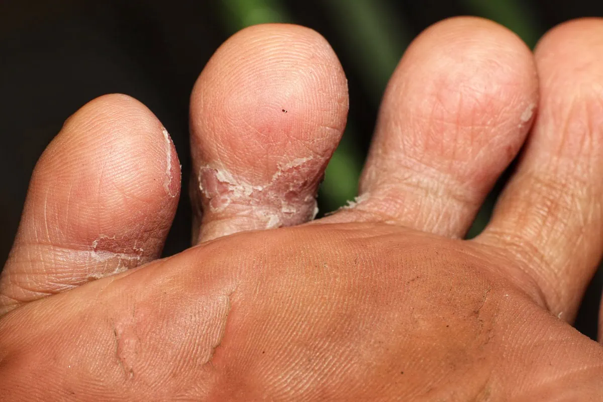
Athlete’s foot is one of the more common conditions of skin of the foot. It is suggested that about 33% to 50% of the population have athlete’s foot at any given time, and that up to 70% of us will have athlete’s foot. Another term for athlete’s foot is tinea pedis referring to it being on your feet. However, it can also affect other parts of your body as it is one of the most common infections of skin. In the feet, it may affect skin and nails. For some patients it may be present as interdigital tinea pedis which means that the moist areas between the toes is where one gets a significant amount of fungal growth.

What are the symptoms of athlete’s foot?

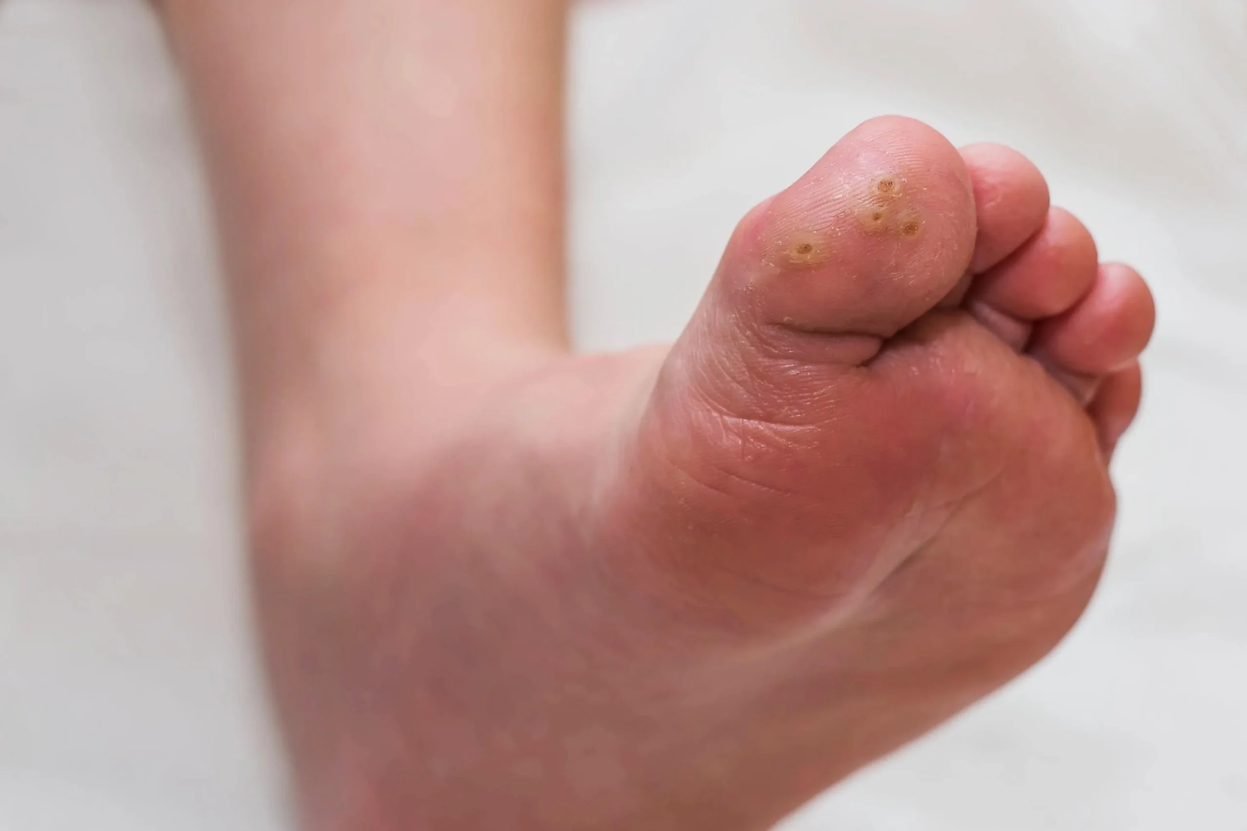
The symptoms can include dry, scaly skin, as well as itching redness and cracking that may become painful in some more severe cases. Typically, tinea pedis presents in a moccasin pike presentation on the bottoms and side of your feet. It may also appear as a vesicular type of infection which means there are small fluid-filled blisters that develop on your feet and they may possibly leak. They may also appear as an ulcerative infection which commonly appears between the toes and may create open sores (ulcers).

What causes tinea pedis?

Fungus likes to grow in warm, moist places, and that is the environment our feet are in most of the time. This makes our feet more prone to contracting or developing tinea pedis. It may also be related to your immune system, and in some situations, because of poor health diabetes as an example, it may make you more susceptible to tinea pedis. For others, however, there may be a part of their immune system that is not able to fight infection on the skin adequately. It is also important to consider that tinea pedis is contagious, especially when you are in public places. It is important not to walk barefoot when you are in public places like community pools.

What are the available treatment options?

Patient education is an important part of learning how to deal with tinea pedis. If you are aware have tinea pedis you may want to consider the following:

  • Drying your feet – after showering or swimming, consider the importance of drying your feet. Also, wash your feet well to keep them clean. Allow your shoes to dry for 24hrs between use
  • Shoes – Avoid rubber or synthetic shoes as these may not allow breathability for the feet
  • Socks – It is important to wear socks that can absorb moisture and keep the feet dry.
  • Going barefoot – if you are not in a public or community area, it is an option to remove your shoes and socks to let your feet dry.
  • Topical treatments for fungal nail infection – there are multiple over-the-counter options that you may want to consider and if you elect to do this, they will usually be applied twice a day because your skin recycles every four to five weeks. Antifungal powder should be considered and used.
  • Lifestyle – It is very important to consider an overall good diet can make your immune system less susceptible to acquiring tinea pedis. Some basic guidelines are to avoid a lot of sugars processed foods and carbohydrates.

Examination

Based upon the examination of your skin and toenails, the doctor may be able to make a diagnosis of tinea pedis. However, some may elect to do a test where potassium hydroxide is used along with scraping of some of the scaly skin to see if the tests come back positive for the fungus.

Treatments for fungal infection of the feet

Topical treatment might include the use of over-the-counter medications and powders

  • Soaks – There are also different types of soaks you can use, one is tea soaks that contains tannic acid which is a very good drying agent. By soaking your feet in this mixture with water for two to three times a week.
  • Lotrisone – Lotrisone is a topical that may be suggested by your doctor if you are experiencing itching with your tinea pedis. It contains lotrimin which is an antifungal and a cortisone cream to reduce itching.

Oral antifungal medication for tinea pedis treatment

A common oral medication that was used years ago is called griseofulvin. In recent times, oral Lamisil (terbinafine) has replaced griseofulvin as the drug of choice. In a study done by Bell-Syer published in Chochrane Database, the efficacy of lamisil exceeded that of the griseofulvin.

  • Risks of taking oral medication -although oral medication may be highly effective, there is the potential of developing liver damage in taking the medication. It is a standard good practice to have a liver panel test done to evaluate the health of your liver before the oral medication is prescribed and to also recheck during the course of treatment. Side effects can be severe, and death is one possible side effect.

How soon do tinea pedis go away?

Tinea pedis often resolve within 2-8 weeks if given proper treatment.

Toenail fungus

Toenail fungus is very common in those who have athlete’s foot. If you have fungus on your skin, the likelihood of it getting into your nails is very likely. The yellow discoloration, and thickened sometimes brittle toenails may become painful and hard to trim

Fungus on the skin can be treated with similar topicals, but these are more likely to be a solution (liquid) rather than a cream. A doctor may also elect to consider the same oral medications for the skin and toenails that were recommended. Again, be advised of the risk of oral medication.

Laser Treatment

Many patients have had the rewarding experience of reversing their toenail fungus with the use of lasers by laser treatment.

How does the laser work for tinea pedis treatment?

For approximately 18 years, we have been treating fungal infections with Pin-point Laser System which delivers rapid pulses of heat into the nail. The heat destroys the fungus, and it is a non-painful treatment and usually takes at least three treatments. Many of our patients have experienced a reversal of their achy-looking toenails with our laser treatment options when all other treatment options have failed.

Lastly, if you have tinea pedis it is important to consider the treatment options you can do on your own and the preventative measures and then seek the medical attention of your podiatrist if these options fail. You may also want to consider laser treatment if you are suffering from toenail fungus. Oral medications in treating tinea pedis should only be considered as a last resort in consideration of the risks that accompany the use of this oral medication.

Tired of the itch and discomfort of Athlete’s Foot? Get lasting relief with expert care at Anderson Podiatry Center. Call now to schedule an appointment in Fort Collins or Broomfield and take the first step toward healthier feet!

Swollen Legs and Edema: Causes, Symptoms, and Treatment Explained

edema in legsIf you note swelling in the lower extremity, the swelling is commonly called edema. Edema is a problem that can be seen in any body part including the abdomen, and arms but it is most common in the feet and lower extremities. Edema in the legs simply means the accumulation of fluid in the body’s tissues. There can be multiple causes and the older you get, the more likely you may experience edema in the lower extremity. It is more common over the age of 65.

What are the symptoms of edema in legs?

  • Tightness or uncomfortable feelings – Because of the pressure of the swelling in the legs, they may feel heavy and may cause dull pain.
  • Difficulty with walking – The swelling may be significant enough that the heaviness of the edema may increase the weight in the limb and make moving the joints more difficult. Walking may be more difficult because in the legs, and ankles, there may be increased weight from the fluid accumulation in the legs or in the feet and ankles and reduction of motion in the joints.
  • Dermatological – The skin may become more stretched and pitted which means when you press on the skin an indentation may remain for a period of seconds. Also, for patients whose swelling is from varicose veins, a dermatological called stasis dermatitis may occur causing a brownish discoloration and itching on the skin.
  • Location – Edema does not tend to be in a small swollen area but rather over a larger region.

What causes edema in legs?

The causes of edema can be multiple and can be related to various medical conditions.

  1. Salt intake – Having too much salt in your diet can cause retention of fluid in the body and this can lead to fluid buildup in the lower extremity.
  2. Venous disorders – The veins are blood vessels that return blood to the heart after it has traveled to the lower extremity via the arteries. People may have had deep vein thrombosis which is a medical term for blood clot. Afterwards, they may have edema in the lower extremity. During a blood clot, swelling may be an early warning sign that it is occurring and if chest pain is associated with this, consider it a medical emergency and seek immediate treatment.
  3. Heart failure– Patients with heart failure are also more likely to have edema in the lower extremity and can be an early warning sign of this condition.
  4. Kidney disease – Kidney disease can also be a cause of edema and may be an early indicator that you may have this problem. If you have swelling in your legs and feet and this is accompanied with difficulty breathing, it would be important to consider kidney disease, call 911.
  5. Lymph nodes – Lymph nodes are part of the lymphatic system and may facilitate drainage from the lower extremity. Some may suffer from lymphedema which causes generalized swelling of both legs in the lower extremity, and it can make walking difficult, and it may cause tightness in the lower legs.
  6. Medication – The side effects of some medications can cause swelling, and this can include medications for high blood pressure, estrogen, and testosterone medications, some medications that treat nerve pain, and diabetic medication.

Treatment

Treatment options for edema in legs include the following:

  • Monitor your salt intake. If the swelling is mild and you believe you have too much salt intake, one of the first steps is to lower your salt intake in your diet.
  • Avoid sitting or standing for long periods of time and instead consider walking or elevating your legs when you’re sitting.’
  • Wear a support hose. Support hose can typically be purchased over the counter and create compression on the limb that will help reduce the swelling. It is important to put this on early in the morning before you walk and when your edema is at its lowest.
  • Treatment of related medical conditions. When the edema is chronic and more severe, it is very important that you see a healthcare provider to rule out problems such as heart failure, pulmonary (lung) problems, kidney disorders, and problems in the circulatory system including the veins and the lymphatic channels.

Treating the cause of the problem

Once the potential cause of the problem is addressed, sometimes medications to control high blood pressure as an example or surgical procedures to address problems such as varicose veins or lymphedema, the swelling may be improved.

The information in this blog should offer you important information regarding the edema in legs that you might be experiencing. One of the biggest takeaways is that edema can be the first warning sign that you are starting to experience a serious issue such as heart or kidney problems. You should not forget that swelling in the legs accompanied with difficulty breathing should be considered as a medical emergency and can be a sign of a blood clot. A quick emergency evaluation is important. Finally, if your symptoms are more chronic and severe, always err on the side of seeking medical attention.

Swollen, heavy legs from edema? Our specialists offer advanced treatments to reduce discomfort and improve circulation. 

Call us today at our Fort Collins location (970) 484-4620, Broomfield location (720) 259-5053 or use our online scheduling system to book your appointment.

High Syndesmotic Ankle Sprain vs. Regular Ankle Sprain: Key Differences Every Athlete Should Know

syndesmotic ankle sprain

Ankle sprains are the most common injury in the foot and ankle. In this blog, we will discuss the two types of ankle sprains. The most common type is a low ankle sprain and a sprain that occurs less frequently is referred to as a syndesmotic ankle sprain or high ankle sprain. You will learn how both these injuries can occur and gain insight into determining if you have such a sprain.

The Basics

Before categorizing a high ankle sprain versus a low ankle sprain, we must first understand the anatomy. The ankle joint is formed by the talus which sits between the two leg bones called the tibial bone and the fibula bone. The fibula is a smaller bone that lies on the outside of the leg and opposite of the other leg.

Low Ankle Sprains

When you have a low ankle sprain, it involves the ligaments that attach the end of the fibula bone on the outside of your ankle and the end of the tibial bone in the inside of your ankle to your foot. There is one major ligament on the inside that connects the tibia to the foot which is called the deltoid ligament. There are three ligaments on the outside of the ankle connecting the fibula to the heel (calcaneus) and the talus bone. These are called the anterior talofibular ligament, calcaneofibular ligament, and posterior talofibular ligament. These ligaments are all involved in low ankle sprains.

High Ankle Sprains

The other ligaments that are referred to as high ankle ligaments support the ankle joint structure are less frequently injured and they connect the leg bones, tibia and fibula, together. When these are injured, it is referred to as a syndesmotic ankle sprain. They are called the anterior and inferior tibiofibular ligament, posterior and inferior tibiofibular ligament, inferior transverse ligament, and the interosseus membrane.

  • Anterior and inferior tibiofibular ligament – this ligament is towards the front of the leg just above the ankle and connects the larger tibia bone to the smaller fibula bone.
  • Posterior and inferior tibiofibular ligament – this ligament connects the back portion of the larger tibia bone to the smaller fibula bone.
  • Interosseous membrane – this fibrous membrane lies between the two ligaments and its function is to connect the tibia and fibula together.
  • Inferior transverse ligament – it goes on the back of the ankle, and it goes from the fibula to the tibia, and it is closer to the ankle ligament.

syndesmotic high ankle sprain

How does a high ankle sprain occur?

High ankle sprain occurs typically when the foot is flexed upwards, and the foot is planted on the ground and there is significant twisting of the leg around the ankle joint most commonly with external rotation. This can put stress on the bones and partially or fully tear the ligaments that are higher above the ankle joint. In contrast, the low ankle sprains are much more frequent with the foot being typically in a downward position, and the foot will rotate inward or outward in relationship to the leg. Syndesmotic ankle sprains occur more frequently in activities such as football where the foot is planted on the ground and possibly because of a collision, twisting is occurring in the leg.

How do I know if I have a high ankle sprain?

Typically, a high ankle sprain will not present with a significant swelling in the outside or inside of the ankle or foot, but pain will be above the ankle joint. You will have swelling in the lower leg region. And also, you may have had the injury when your foot was planted firmly on the ground and the foot was flexed upward.

How is a syndesmotic ankle sprain diagnosed?

The doctor will inquire about how the injury occurred. The examination will usually show pain and swelling in the lower leg close to the ankle and the doctor will perform a squeeze test where the tibia and fibula bones are squeezed together, and this will typically create pain.

X-rays

X-rays will be taken, and, in some cases, there will be a fracture noted particularly in the fibula bone or there could be a portion of bone that was pulled away by one of the injured ligaments. If there appears to be a separation between the tibia and fibula bone on x-ray, surgery may be needed to bring the two bones back into a more anatomical alignment.

CAT scans

CAT scans may be useful to more specifically evaluate for possible bone injury to the tibia and fibula and to ensure that no fracture is associated with syndesmotic injury.

Treatment for a syndesmotic ankle sprain

  • Rest, Ice, and Compression – if the injury is milder, cutting back on activities and resting the area, icing, and wrapping it with compression is helpful. Anti-inflammatory may be used
  • Physical Therapy – physical therapy may be needed to reduce the swelling and promote returning to full activity.
  • Immobilization – in more severe cases, it may be recommended not to bear weight on the ankle for a period of time or simply wear a CAM walker as part of the recovery.
  • Surgery – if there is any fracture where displacement may be present or an injury was severe enough to cause more separation between the tibia and fibula bone, surgery may be suggested.

Because ankle sprains are one of the most common injuries of the lower extremity, it is important to understand the concept that they are frequently under-evaluated and under-treated. For many, treatment of an ankle sprain may involve a visit to an urgent care facility, nothing shows up on an x-ray and they are advised to use an ace wrap, ice the area and gradually return to regular activity.

It is important to consider getting a consultation with a foot and ankle specialist to make sure you get an accurate diagnosis, and that appropriate treatment is done early so that you may get back to full activity sooner with less ankle complications in the future. Finally, syndesmotic ankle sprains are a rare occurrence but important for you to know about so that if you suspect you have one, you can seek a consultation with a foot and ankle specialist.

Call us today at our Fort Collins location (970) 484-4620, Broomfield location (720) 259-5053 or use our online scheduling system to book your appointment.

Complex Regional Pain Syndrome: The Story You’re Not Hearing

Complex Regional Pain Syndrome CRPS

Complex regional pain syndrome (CRPS) is a form of chronic pain that could affect the parts of the body, mostly the arm, leg, hand or foot. It is not fully understood what causes complex regional pain syndrome. By reading this blog you should be better informed if you have this condition as early treatment can be very important. Another term that is often used is called reflex sympathetic dystrophy which has similar symptoms but tends to be a more severe state of complex regional pain syndrome.

The signs and symptoms of Complex Regional Pain include the following

  • Changes in skin temperature – the affected area may be warmer or cooler.
  • Changes in skin color – the skin may be whiter or have a purplish or bluish discoloration.
  • Muscle spasms and weakness in severe cases.
  • Burning and/or throbbing pain.
  • Sensitivity to touch or cold temperatures.
  • Decreased ability to move the affected limb.

It is important to note that occasionally CRPS can spread elsewhere in the body including the opposite limb. The symptoms of CRPS may go away on their own but for many the symptoms may persist for months or years and early treatment is most effective to reverse the symptoms.

Causes of Complex Regional Pain Syndrome

Conventional Medicine categorizes CRPS into two types:

  • Type 1 – this is often referred to as reflex sympathetic dystrophy and may occur after an injury or an illness but there may not be any direct damage to the nerves of the affected area. Most people are thought to be type 1.
  • Type 2 – this is often referred to as causalgia. The symptoms of this type are similar to type 1; however, type 2 occurs after a specific nerve injury.

The following are examples of what may cause the symptoms of CRPS. They include a crushing injury, fracture, ankle sprain, heart attack, strokes, and for others, it may be a result of surgery on the hands, feet, or legs. With more chronic CRPS, treatments may be more challenging as it may start to affect the central nervous system also. Whereas in the beginning, it was a localized issue in the lower extremity. The full mechanism between the central and peripheral nervous systems is not well understood as it relates to CRPS.

How do doctors diagnose CRPS?

  • Patient’s History – The patient will report an illness, injury, or surgery where symptoms started for many people.
  • Specific tests to diagnose CRPS include Ultrasound or Magnetic Resonance Imaging (MRI) which may be used to identify underlying nerve damage.
  • EMG may also be used for testing nerve function.

Conventional Treatment Options

For many, the treatment of CRPS involves seeing a doctor who specializes in pain management, and the pain management doctors may use the following treatments.

  • Physical Therapy/Occupational Therapy
  • Psychological and Behavioral Therapy
  • Medications – this can include nonsteroidal anti-inflammatory drugs, anti-depressant drugs that may be used, and also anti-seizure drugs such as gabapentin and pregabalin. These medications are usually prescribed to treat CRPS.
  • Narcotics – If other medications fail, narcotics may be used and carefully monitored by the prescribing doctor.
  • Topicals – topical analgesic creams can be used such as lidocaine.
  • For severe symptoms, the use of spinal cord stimulation.
  • Sympathetic nerve blocks – these blocks involve injections to the lumbar area of the back which can offer pain relief but is usually temporary.
  • IV ketamine infusions – this is another treatment option.

Things that you can do at Home

  • Exercising on a daily basis is important to move the affected body part and it may help repair the damaged nerves.
  • If there is swelling present, compression stockings may also help.
  • If there is a swelling in the limb, elevation while resting may also be helpful.

The game-changing approach that peripheral nerve surgeons are using to discover the root cause and reverse CRPS.

The Non-conventional Treatment Options

Demystifying the cause of Complex Regional Pain Syndrome: Why you should consider consulting with a peripheral nerve surgeon

With the advancement of the knowledge of the peripheral nerve system in the lower extremities, a subspeciality of doctors has been created that specializes in the evaluation and treatment of peripheral nerve disorders including neuropathy, restless legs, and chronic pain. From our knowledge and experience, we are now able to reverse the symptoms of CRPS by evaluating and treating the injured nerve. This has helped many patients avoid the need for spinal cord stimulators and has allowed others to get off narcotics.

What are the options a peripheral nerve surgeon would offer?

  1. Injections – Injections may be used in the early stages to reverse the symptoms of the damaged nerve but are also helpful in identifying which nerve branches are damaged.
  2. Laser Treatments and Electrical Stimulation Treatments – These treatment modalities can be used if in mild cases.
  3. Surgery– Surgery may involve the removal of the damaged nerve branch and also burying the nerve in the muscle to prevent regrowth.

The goal of a peripheral nerve surgeon is to discover the specific nerve that has been damaged and address treatment locally in the foot or leg. Thus, eliminating the need for medication or spinal cord stimulators for many. If you would like to learn more about these options, please consider visiting https://www.aens.us/aens/default.asp

Call us today at our Fort Collins location (970) 484-4620, Broomfield location (720) 259-5053 or use our online scheduling system to book your appointment.

A New Cause of Muscle Twitching in Legs and a Solution

muscle twitching in legs restless legs syndrome
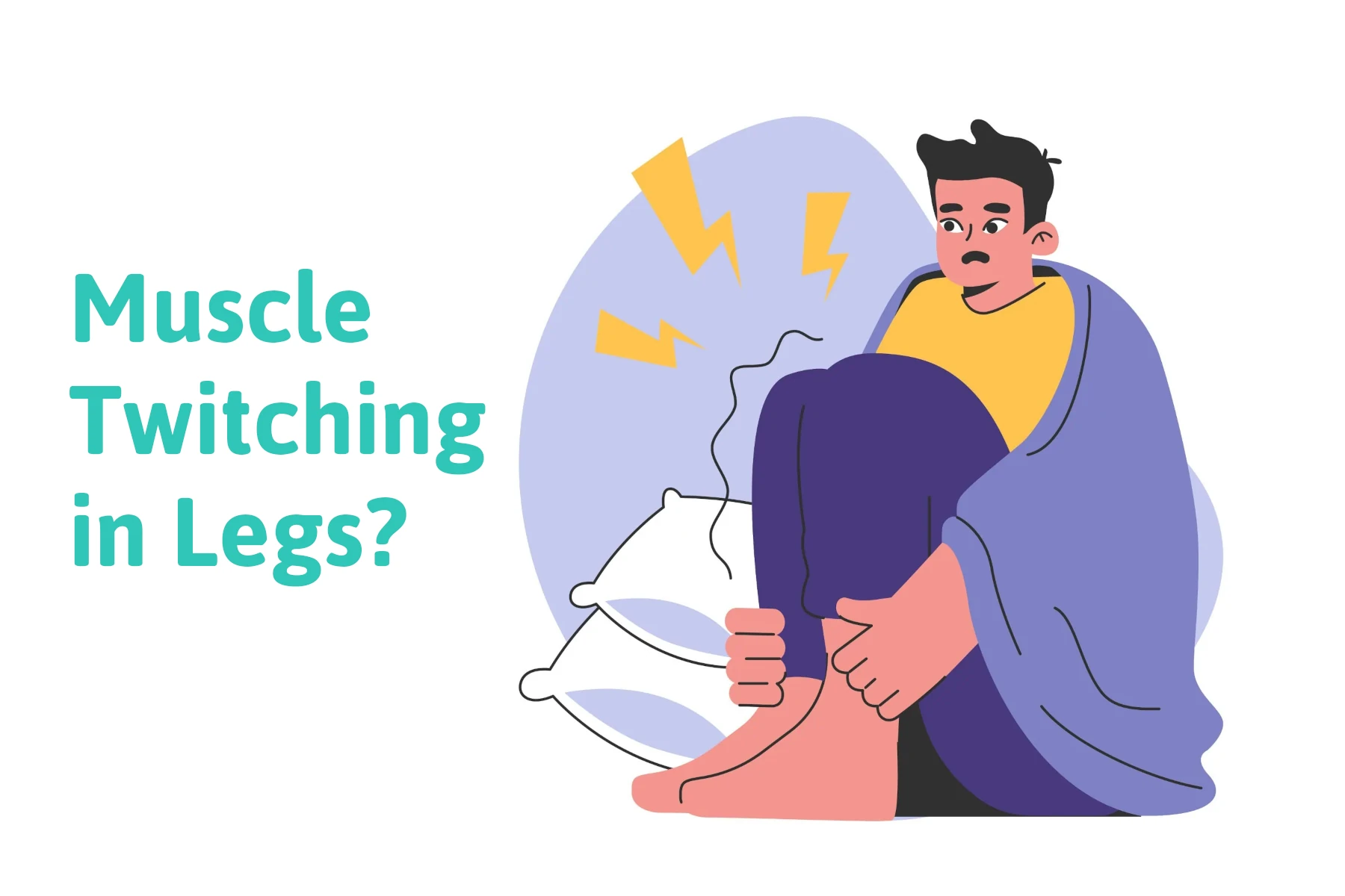
Twitches are common. Muscle twitching is a common complaint that can be associated with restless legs (RLS). You do not have to have restless legs to have muscle twitching, but it can be a symptom related to restless legs that is annoying, and in its more severe forms can present as jerking in the legs. Another term to describe this is periodic limb movement disorder (PLMD). Another common medical term to describe muscle twitching is muscle fasciculations. Both muscle twitching and muscle fasciculations can be used interchangeably. Muscle twitching can be in different parts of the body. In this blog, we will specifically focus on muscle twitching and how it relates to restless legs syndrome but before we do so we need to mention other medical conditions or reasons that you may be getting muscle twitching in your leg.
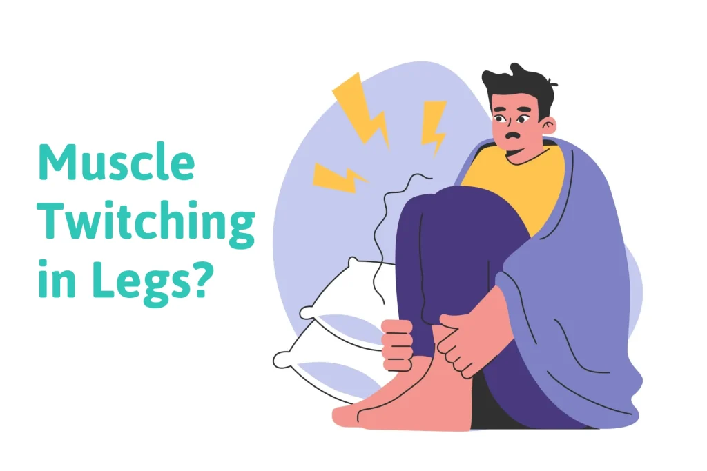
  1. Amyotrophic Lateral Sclerosis (ALS), or also known as Lou Gehrig’s Disease – This is a severe neurological condition that includes speech difficulties such as slurred speech and difficulty speaking. It can also cause muscle weakness that can become severe in the hands, arms, feet, and legs. If you are having significant weakness of muscles or difficulty speaking, this could lead you to suspect ALS.
  2. Increased adrenaline – Anything that stimulates the body such as caffeine or stress may also be a cause of muscle twitching.
  3. Fatigue – Any type of activity that may cause muscle fatigue may result in muscle twitching along with muscle spasms and muscle cramps after the activity is completed.
  4. Benign Fasciculation Syndrome – This is a medical condition that can cause fasciculations in various muscle groups of which there is no known cause.

Treatment of muscle twitching in legs

Because these symptoms are not serious, there aren’t any FDA-approved treatments that are known to stop muscle twitching. There may be medications that are used to relax the muscles and reduce muscle twitching. However, the side effects of these medications are not worth the benefits.

Muscle Twitching/Jerking as it relates to Restless Legs

A New Hypothesis

To understand muscle twitching as it relates to restless legs, we first must understand the two basic anatomical components of the nervous system.

Central Nervous System

The central nervous system is composed of the brain and the spinal cord. Most research regarding restless legs involves the central nervous system. It is thought that lower dopamine levels or lower iron levels in the brain may correlate to having restless legs. Oftentimes medications are used to increase dopamine levels or iron levels in the brain.

Peripheral nervous system

The peripheral nervous system is composed of nerves that extend to the arms and legs. An example in the upper extremity is the median nerve in the wrist which undergoes compression as it passes through a tunnel called the carpal tunnel. Another area is the ulnar nerve in the elbow which is also referred to as the funny bone when you hit your elbow. This new idea about jerking or twitching is that it may be caused by damage to the nerves of the peripheral nervous system.

Background

This author has successively reversed the symptoms of neuropathy for over two decades. The symptoms of neuropathy are reversed by opening (decompressing) different nerve tunnels in the lower extremity. By releasing the compression on these nerves, the symptoms of neuropathy can be improved. Approximately ten years ago, it was found that two additional nerve tunnels could be causing nerve compression and seem to be highly correlated to restless legs. By relieving pressure on the nerves in these two tunnels, reversal of restless legs symptoms was possible. Not only were the symptoms of nervousness, restlessness, creepy crawly sensations, and cramping released but also jerking and fasciculations were also reduced or eliminated.

How This Concept Disrupts Conventional Medicines Theory on Twitching and Jerking related to Restless legs?

Until the recent observation of patients having great success with nerve decompression surgery helping restless legs, it had always been assumed that iron and dopamine may play a major role. That is not to say that they may have some impact, however, it is important to note that surgical decompression has shown success and should be considered an option especially when restless legs symptoms are severe.

The new hypothesis for muscle twitching in the leg.

The idea is that the twitching can be caused by the peripheral nervous system when the nerves are damaged from excessive compression. This damage to the nerve may cause a noxious sensation in the legs and send a signal to the spinal cord and the central nervous system that results in a twitching or jerking response in the muscles in the leg.

In some cases, severe jerking is involuntary meaning it cannot be controlled by the patient. Another possibility is that the compression also damages the motor neurons that stimulate muscle contraction. This may also create twitching and lead to muscle weakness which is seen in some patients with restless legs. With the noxious stimuli, if it is mild, the patient may present with fasciculations. Currently, we have one peer-reviewed published paper showing the fact that we can reverse restless legs symptoms, and another paper soon to be published. Both papers report that jerking is eliminated or greatly reduced.

So, if you have severe symptoms of restless legs that are affecting your sleep and also there is twitching in your legs, understand that there may be another option. This new option of decompression of tight nerve tunnels in the legs tells us there’s a possibility that the twitching may be related to the compression of the nerve. I often refer to restless legs as restless legs compression syndrome as for many there is compression in the nerve tunnels in the legs where the symptoms of restless legs are found.

Experiencing unexplained muscle twitching in your legs? Our foot and nerve specialists can help pinpoint the cause and find a solution.

Call us today at our Fort Collins location (970) 484-4620, Broomfield location (720) 259-5053 or use our online scheduling system to book your appointment.


David Post Operation

Arlene is cured of muscle twitching in her legs after 25 years

Do You Have Stinky, Itchy Feet? You May Have Pitted Keratolysis

pitted keratolysis

If you have feet that are very itchy and have a bad odor, chances are you might have pitted keratolysis, also known as keratolysis plantare sulcatum. The foot is an area that can perspire quite a bit and might put you at risk of acquiring pitted keratolysis. Topical antibiotics are frequently part of the treatment option that a podiatrist may suggest to get rid of this disorder.

Who is affected by pitted keratolysis?

  • It tends to be more common among men.
  • Feet that perspire too much (hyperhidrosis).
  • People that live in warm, humid climates.
  • People that are on their feet working in hot climates.
  • How do I know if I have Pitted Keratolysis?

Location

Typically, the skin disorder will present on the soles of the foot, the pressure-bearing plantar surface areas. These weight-bearing areas including the ball of the foot is where a lot of perspiration takes place and may be where most of the bacteria may preside. The condition may also be in the web spaces in between the toes. An odor will be present. The unpleasant foot odor associated with pitted keratolysis is from sulfur compounds produced by bacteria.

Appearance

It usually presents as a white patch on the skin that is lighter than your natural skin color and will have pits that look like small holes or indentations in those locations. Oftentimes these small holes (pits) will cluster together and may cause a sore. Because of the clustering of the pits, it may be referred to as ringed keratolysis. There will also be an itchiness in the skin that is affected.

Bacteria that cause pitted keratolysis

The most common bacteria that cause pitted keratolysis are:

  • Dermatophilus congolensis
  • Kytococcus sedentarius
  • Streptomyces
  • Corynebacteria
  • Trichomycosis axillaris presents in the armpits and is caused by corynebacteria. The armpits, much like the feet, are warm, moist areas and the corynebacteria can cause both conditions.
  • Actinomyces

The one thing all these bacteria have in common is they like to breed in warm, moist conditions. The bacteria will then proliferate and produce an enzyme. These bacteria also have a protease enzyme that can destroy skin cells going down into a layer of the skin called the stratum corneum, also known as the horney layer.

How is the diagnosis made?

The presentation of pitted keratolysis will help to put this skin condition in a differential diagnosis that may include other dermatological conditions by doing a culture test, taking a swab of the skin or scraping a small amount of the infected skin. The podiatrist can assess if one of the bacteria that causes these disorders is present in the culture. A small skin biopsy can also be done where a small skin tissue can be looked at with a microscope. Another means of diagnosing pitted keratolysis is using coral red fluorescence. Under a Wood Lamp, the corynebacteria will show coral red fluorescence.

Differential Diagnosis of athlete’s foot and pitted keratolysis

Athlete’s foot is a very common foot complaint and more common than pitted keratolysis. Patients may think they have athlete’s foot but the difference between the two is pitted keratolysis is a bacterial infection whereas athlete’s foot is from a fungal infection. Both can cause itching, and both are present on the bottom of the feet. Pitted keratolysis has a different appearance than athlete’s foot as it has pitting on the skin while athlete’s foot has scaling of the skin. Also, pitted keratolysis causes a significant odor whereas athlete’s foot usually does not.

What you can do at home?

If you believe you have pitted keratolysis, it is important to wear shoes and socks to breathe. This will allow the reduction of perspiration that will stay on your skin. You may also want to consider antiperspirants that you can buy over the counter if you sweat a lot. Washing your hands and feet when you remove your shoes and socks should be done, and you can also change your shoes and socks when they get wet.

Home treatments may include essential oils such as tea tree oil and also soak your feet in a diluted mixture of hydrogen peroxide and water. The ratio should be 1 part hydrogen peroxide and 3 part water and the soak should last for a couple of minutes.

What treatment options would your podiatrist suggest?

Topical Antibiotics

After doing appropriate testing to identify which bacteria is affecting your skin, there are several topical antibiotics that can be placed on the skin as part of the treatment of pitted keratolysis and this includes topicals that can contain erythromycin, clindamycin, mupirocin, or fusidic acid.

Antiseptics

Benzoyl peroxide comes in a gel or cream form and can also be applied to the skin to help fight the skin infection.

Injections

Botulinum toxin injections can be used and are injected locally in the area of the skin condition when the pitted keratolysis does not respond to the other treatments.

Treating Excessive Sweating

Excessive sweating refers to hyperhidrosis and your podiatrist may prescribe antiperspirants so that the condition does not return.

When should I see my podiatrist?

If you have done the suggested home and the problem persists, you should seek treatment from your foot specialist. This diagnosis can be difficult to treat and for many, this is a dermatological condition where you would likely need the help of a healthcare professional to address it.

Call us today at our Fort Collins location (970) 484-4620, Broomfield location (720) 259-5053 or use our online scheduling system to book your appointment.

Understanding Stress Fractures in the Foot: Symptoms, Causes, and Prevention

stress fracture in foot

Stress fractures are one of the common athletic injuries in the foot and ankle. It has been said that they may comprise 20% of these injuries. A stress fracture is caused by putting too much stress through a bone that does not have time to recover. At first, it may be considered a bone bruise but with repetitive stress, a break may develop in the bone. By definition, a stress fracture is not typically from one traumatic episode. The challenge of a stress fracture in the foot is that the diagnosis could be missed, and treatment could be delayed. The message of this blog is that the earlier a stress fracture is detected, the faster you’ll receive treatment. Thus providing you with the opportunity to get back to regular activity faster.

Symptoms of a Stress Fracture: How do you know if you have a fracture in your foot?

Generally, no matter where a stress fracture is occurring there tends to be swelling around the injured bone. Typically there will be a high amount of pinpoint pain directly in that area. Usually pressing directly on the bone will cause pain. The pain associated with the stress fracture may make walking difficult!

Just because you can walk on your foot does not mean you don’t have a fracture.

The most common location for stress fractures in the foot tends to be the metatarsal bones. These are the long bones that start in the mid-foot area and extend down to the toes. Ending with the weight-bearing bones of the ball of your foot.

Risk Factors: How Does a Stress Fracture Occur?

High Impact Activities

High-impact activities are common causes of stress fractures. These could include sports such as track and field, running, or other high-impact sports such as tennis, basketball, and football. All of these are repetitive activities that put added stress on the foot and ankle which can lead to a stress fracture.

Improper training can lead to stress fractures. Doing too much too soon or not having rest days between the high-impact activities will not allow your body time to repair. It is important to have rest days for the body to rest or you may develop stress fractures.

Foot Types

Some foot types are more prone to stress fractures. Some people are born with a shorter 1st metatarsal, and this is the bone that makes up the great toe joint. If this bone is excessively short, this will cause more weight bearing on the 2nd and 3rd metatarsal bones.

Bone Insufficiency

Fractures are more common with the following:

Hyperparathyroidism

If the parathyroid gland is hyperactive, the parathyroid hormone will take calcium from the bone and put it in the bloodstream thereby creating weakness in the bones.

Osteoporosis

Osteoporosis appears more frequently in women than in men and it could be related to eating disorders or menstrual dysfunction.

Vitamin D

Vitamin D deficiency can also cause weakness of bone and can be more common in winter months when sun exposure is more limited.

Diagnosing a Stress Fracture

When you see a foot specialist, they will examine your foot and look for swelling and pain. The clinical exam itself is important and will lead the podiatrist to suspect a fracture.

X-rays

X-rays are taken but stress fractures may not be seen if taken soon after the symptoms have started. Oftentimes, the fracture is so small that it is not seen on X-rays. Ten days after the fracture starts, new bone will form that is visible on x-ray and this is called a bone callus. That is why X-rays may show no signs of a stress fracture but if the x-ray is repeated ten days later, the bone callus will be seen, and the diagnosis can be made.

MRI – Magnetic Resonance Imaging MRI

This can be considered the gold standard and is highly recommended especially if x-rays are negative for a stress fracture, but the symptoms correlate to a stress fracture.

Bone Scan

A bone scan can also be performed which will more accurately detect a stress fracture than x-rays.

Treatment

With very few exceptions, the use of a CAM walker is recommended. For many people, they can continue weight bearing in a CAM walker. The use of a CAM walker can last from 3 to 6 weeks depending on the severity of the stress fracture. In the rare instance that the bone is displaced at the fracture site, this may necessitate the need for non-weight bearing before going to weight bearing in the CAM boot. The reason for this is to prevent further displacement of the bone.

cam walker foot stress fracture in foot

Location of Stress Fractures in the Foot

There are many types of stress fractures.

Metatarsal bones

These are the most common bones to have stress fractures especially the 2nd and 3rd metatarsals and to a lesser degree the 4th and 5th metatarsals.

Heel Bone: Calcaneus

This is an uncommon stress fracture. When a stress fracture occurs in the heel, it is usually quick to heal because it has a very good supply.

Tarsal bones

There are many cube-shaped bones in the midfoot area where stress fractures may also occur but again are not as frequent as the metatarsal bones.

Sesamoid bones

Two small bones underneath the big toe joint function much like the patella in the knee joint. These occasionally can fracture and can be caused by trauma to the big toe area but less frequently, can be caused by overuse.

In summary, early detection of a stress fracture is vital. So, it is important not to ignore your symptoms, especially if they’re located in the region of the metatarsal bones as early detection is important. It can reduce the time to heal the stress fracture if early treatment is administered.

Call us today at our Fort Collins location (970) 484-4620, Broomfield location (720) 259-5053 or use our online scheduling system to book your appointment.

A New Treatment to Remove the Root Cause of Plantar Warts: SWIFT Laser Device

what are the causes of plantar warts

Plantar warts are small, rough growths that appear on the soles of the feet, caused by an infection from the human papillomavirus (HPV). This is a type of virus that can also cause genital warts, and it can be spread. The virus enters through tiny cuts or breaks in the skin. We’ll also take a closer look at how plantar warts develop. By understanding how they develop, you can take steps to reduce your risk and prevent their spread.

What is the cause of plantar warts?

The causes of plantar warts are simply that your body’s immune system is not fighting the wart virus. Yes, warts are contagious but why do some people get them more than others? This can be from an immune system not strong enough to fight the virus.

  • Weakened Immune System: Individuals with weakened immune systems are more susceptible to developing plantar warts which may correlate with alterations in hormone levels.
  • Hormones: Examples of how hormones may play a role include the tendency for warts to appear more frequently during puberty and a possible association with birth control pills.
  • Direct Contact: Touching a wart or sharing personal items like towels or shoes with someone who has warts can lead to transmission.
  • Moist Environments: HPV thrives in warm, moist environments, making feet particularly vulnerable.
  • Walking Barefoot: Walking barefoot in public areas like locker rooms, swimming pools, or communal showers increases the risk of contracting HPV.

Presentation of warts on your feet

Warts can occur anywhere in the body, but the most common part of the body where they appear is on the feet typically on the bottom of the foot or in the toes. They appear harder than normal skin and can cause pain when pressure is applied to them, especially when squeezed. They oftentimes will have dark specks on them and when trimming a wart, there can be small areas of pinpoint bleeding because there are tiny blood vessels within the wart tissue. A wart can present in a single area and the size can vary greatly. The most difficult presentation of a wart is called a mosaic wart. This type of wart consists of multiple small warts over a large area.

plantar-warts-infographic

Treatments for plantar warts

Home Remedies

The following are home remedies that you may want to try.

  • Salicylic Acid – There are over-the-counter products with salicylic acid, commonly used for treating warts caused by HPV, which is also one of the causes of plantar warts. These products are applied daily and slowly eat away the wart. If the warts are multiple or large, this process may take a while, as over-the-counter salicylic acid is not a prescription strength.
  • Apple Cider Vinegar – Apple cider vinegar is thought to be acidic and can help destroy the wart tissue. The suggested method is applying apple cider vinegar to a cotton ball and taping it to the wart overnight.
  • Pumice Stones – Often used alongside salicylic acid, a pumice stone can be rubbed on the wart to remove dead wart tissue.
  • Duct Tape – Some find that applying duct tape to the wart daily helps to break down the wart tissue.

Conventional Treatments your Podiatrist may recommend

  1. Salicylic Acid (Prescription Strength) – Salicylic acid with a higher concentration or percentage than over-the-counter products may be applied to the foot and reapplied every two weeks. The patient returns to the office and the dead wart tissue will be trimmed away. Salicylic acid will then be reapplied until the wart is gone. The number of treatments can greatly vary.
  2. Cantharone – Cantharone treatment is similar to salicylic acid. However, Catharone creates an allergic reaction (blister) which is destructive to the wart and the treatments are every two weeks until full destruction of the warts has taken place.
  3. Surgical Excision – When surgery is performed, it is usually in a clinical setting and you can walk immediately. Anesthesia is used to numb the area.
  4.  Laser Treatments – Various forms of laser may be used to destroy wart tissue.
  5. Bleomycin Injections – This can be done into the wart tissue as bleomycin injections are thought to be destructive to the wart.
  6. Liquid Nitrogen – Liquid nitrogen can be used to freeze the wart, and it is used a lot by primary care doctors and dermatologists as it is used throughout the body, but it is not favored by most foot specialists as we believe it is not as effective on warts of the foot. The reason for this is that most warts on the foot are on weight-bearing areas of the skin and the body’s weight pushes those warts deeper into the skin where the liquid nitrogen may not penetrate.

The Fallacy of Wart Treatments

All home remedies and conventional treatments by doctors have one thing in common: they are all destructive in nature. The goal is to destroy the wart by different means, but the underlying causes of plantar warts have never been fully addressed until now. Continue reading to understand how the SWIFT device targets the root cause of the problem, helping to reduce the recurrence rate.

The SWIFT device as a treatment for Plantar Warts is a game changer! It can strengthen the immune system to get rid of the wart and once treatment is completed, the patient is less likely ever to have wart recurrence.

Introducing the SWIFT device for the treatment of plantar warts

The SWIFT device was introduced as a medical device approximately 3 years ago and it is the only known treatment that is able to treat the cause of the wart.

How does the SWIFT device work for plantar warts on your feet?

The SWIFT device produces microwave heat energy and delivers microwave heat into the tissue. The treatment involves five two-second applications of this energy. There are no side effects afterward and no discomfort after treatment. This microwave heat has an effect on heat shock proteins within the viral tissue. The heat shock proteins are released during the treatments which alert the immune system of the HPV virus. By altering these heat shock proteins, the body’s immune system recognizes the virus and attacks the wart tissue. A minimum of three treatments are needed spaced at one-month intervals.

The SWIFT device has been our treatment of choice for plantar warts as it provides a safe and effective option. It does not require the patient to have destructive, painful treatments that may require dressings or keeping the foot dry during treatment. It is also the smartest way to treat warts as we’re not managing the condition but rather using a technology that addresses the root cause. So, if you have been struggling with wart treatments, please consider this new technology as the preferred option. With two convenient foot and ankle clinical locations in Fort Collins and Broomfield, Colorado, our experienced doctors will talk you through the procedure and what to expect. Check our YouTube video on plantar warts here.

Call us today at our Fort Collins location (970) 484-4620, Broomfield location (720) 259-5053 or use our online scheduling system to book your appointment.

Frequently Asked Questions About the Causes of Plantar Warts

  • What causes plantar warts to develop?
    Plantar warts are caused by the human papillomavirus (HPV), which infects the outer layer of skin on the soles of your feet.
  •  How does HPV enter the skin to cause plantar warts?
    The virus enters the skin through tiny cuts, abrasions, or cracks on the surface of the feet, often in moist environments like locker rooms or swimming pools.
  • Is HPV contagious, and can it spread to others?
    Yes, HPV is contagious and can spread through direct contact with an infected person or surface, such as shared showers or towels.
  • Why do some people get plantar warts while others don’t?
    Susceptibility depends on individual factors like immune system strength, skin integrity, and frequent exposure to high-risk environments.
  • Can wearing certain types of footwear contribute to plantar warts?
    Yes, wearing shoes that trap moisture or walking barefoot in public places increases the risk of contracting the HPV virus and developing plantar warts.

The Basics of Plantar Fasciitis Stretching and Why It Can Fail You

plantar fasciitis stretching

Pain on the bottom of the heel is possibly the most common foot complaint that a foot specialist sees. There are many options to reduce or reverse these symptoms and the purpose of this blog is to offer stretching options that you may want to consider. The most common cause of heel pain is a problem called plantar fasciitis. The plantar fascia is a tight connective tissue that starts at the ball of the foot and extends to the bottom of your heel. With every step that you take, as the arch flattens, it is thought that the fascia is pulled, and this creates stress where it attaches to the heel. In younger people, the fascia tends to be more elastic and more stretchable, however, as we get older it is thought to lose a lot of its elasticity. The idea is that with a lack of flexibility, it is pulling from your heel, and microscopic tears may be occurring. That is why for many of you searching for solutions to your plantar fasciitis, stretching is brought up as a solution.

This blog intends to introduce you to some basic plantar fasciitis stretching exercises that you may perform. I will also explain why you may want to reconsider this option, especially if you don’t see progress, and also explain why it may make the problem worse and could be a dead-end solution. I will offer scientific reasons for avoiding plantar fasciitis stretching in certain cases.

The basics of the heel pain

There are two things to consider when using stretching to reduce or eliminate heel pain:

  • The plantar fascia. The plantar fascia is a very strong tissue often referred to as a tendon or it has been described as the ligament that connects your heel to the ball of your foot, but it is simply a tight connective tissue (fascia). The theory is if you can stretch it, it will reduce the irritation that is created at the bottom of your heel where it inserts into your heel bone (the calcaneus). So, many stretches are focusing on stretching the fascia specifically.
  • The Achilles tendon. Other stretching exercises are targeted to the calf muscles which include the gastrocnemius muscle which is the large muscle in your calf but also the soleus muscle which is a thinner muscle deep beneath the gastrocnemius muscles. Both of these muscles attach or form the Achilles tendon. If either one of them is tight that will reduce the amount of upward motion at your ankle joint, this is called dorsiflexion. If you have tightness in the calf area (the Achilles tendon), then that lack of upward mobility at the ankle will cause more strain on your fascia. If you have an extremely tight Achilles tendon, your heel will lift off the ground earlier as you propel forward because of restricted motion at your ankle therefore causing more pull on the fascia. So, stretching the calf muscles is thought to be helpful.

Stretching the plantar fascia

  1. Stretch On a Step. Using stairs is a useful exercise to stretch both the fascia and the Achilles tendon. To do this stretch, stand facing the stairs with the ball of both feet on the edge of the step and the heel not in contact with the step. You can do this stretch with both feet or with one foot at a time and you may need to use the handrail if you are using one foot. The idea is with your knee extended and not flexed, allow your heel to drop down and this can provide stretching for the fascia and the Achilles tendon. You could do this with 15 repetitions per side two times a day.
  2. Frozen Water Bottle Stretch. This stretch involves using a frozen water bottle that you put on the floor and while you sit in a chair you gently roll the water bottle on the arch of your foot. Using the ice in the water bottle and the rolling technique may gently pull on the fascia to stretch it and reduce inflammation of the fascia.
  3. Standing Calf Stretches Against a Wall. While standing facing a wall, put both hands on the wall, and with the foot needing to be stretched furthest away from the wall, lean forward with the affected leg straight so that you are not bending at the knee and do this until you feel a stretch. The opposite knee will be flexing as you lean forward towards the wall. It is important to use proper technique when doing this by keeping your knee straight it creates more stretch of the gastrocnemius muscles compared to the knee being flexed. Also, your foot must be facing directly straight and not pointing outwards because with it pointing outwards, there may be more tendency for the arch to flex or bend thus reducing the stretch in your calf area.

Exercises for plantar fasciitis

When seated in a chair, place a towel on the floor and use your toes to grab the towel doing multiple repetitions. This is thought to reduce stress on the fascia by increasing strength in the muscles that form your arch.

Current science to challenge the reasoning behind plantar fasciitis stretching exercises being the solution

Why does healthcare continue to claim that the fascia is inflamed and keep using the term plantar fasciitis when this has been disproven?

Several years ago, Dr. Harvey Lemont did a study where he studied the plantar fascial tissue for evaluation when he did surgery on 50 subjects. Only one of those specimens showed the fascia was inflamed and 49 showed that the fascia was tissue that was breaking down (plantar fasciosis). This supports the idea that microscopic tears are taking place and supports the fact that the use of human cellular tissue products are far superior treatment than using anti-inflammatories such as oral medications or cortisone injections. This would also argue that stretching may not be a good idea as you’re damaging the tissues even more and causing more tearing. Also studies out in Europe support the success of regenerative medicine versus cortisone injections. Our patients appreciate the success they’ve had with the stem cell options.

Patient Testimonial

Orthotic Devices for plantar fasciitis

Orthotic devices have been used as one of the primary solutions for chronic heel pain for foot and ankle specialists for many years. The orthotics do nothing to stretch the fascia but in fact reduce the pulling of the fascia from the heel.

In summary, it has been this doctor’s experience that plantar fasciitis stretching can be a bad idea. It may give you some relief but does not reach the point of actually solving the problem. People simply manage the condition and this may greatly limit what they do. It never gets them to fully resolve it. Finally, our primary goal is to permanently resolve the problem, and we have found that the best strategy is to structurally reduce the pull of the fascia from the heel. The evidence is there to support that microscopic tears occur while stretching. Using human cellular tissue products (regenerative medicine) to repair the damage brought on by the tearing of the fascia can reverse the damage. So, consumers beware! If you are frustrated with how little stretching is helping you or it is making it worse, consider the treatment that we offer that is more science-based.

Call us today at our Fort Collins location (970) 484-4620, Broomfield location (720) 259-5053 or use our online scheduling system to book your appointment.

Stop Putting Up with Your Leg Pain and Treat the Cause!

causes of leg pain

Leg pain is a common complaint that can include a multitude of causes. The purpose of this blog is to give you the more common causes so that you may better understand your leg pain. Generally speaking, leg pain can be caused by injury, overuse, and also from general health conditions. It can also be related to circulatory problems and nerve conditions. This blog may serve as a general guide to know what type of specialist you should see that can treat leg pain based upon which of the following symptoms you may have.

Circulatory conditions to be aware of for causes of leg pain

Circulatory conditions that may cause pain in the legs include varicose veins, thrombophlebitis (blood clot), and peripheral arterial disease.

  • Varicose Veins – This is a common condition caused by veins that do not function properly. Arterial blood flows down the extremity to the foot and has to travel back to the heart through the veins. There are one-way valves in the veins that oftentimes work improperly, and their function is to not allow the backflow of venous blood back toward the foot. When these veins are dysfunctional this can lead to a buildup of venous blood in the veins causing them to enlarge. These are called varicose veins and they can be unsightly and painful.
  • Thrombophlebitis (Blood clot) – A blood clot may occur when the blood in the vein traveling back up to the heart becomes clotted. This can happen in the superficial veins in the lower leg closer to the skin and generally will cause pain in the area of that vein. As the veins travel back up towards the heart, they travel deeper into the leg, especially when they approach the knee joint area. If the deeper veins become blocked with a blood clot this is called deep vein thrombosis (DVT). This will cause more generalized pain in a larger area and possibly the entire leg and it is critical you seek medical attention. If the blood clot breaks off and travels to the heart and lungs, this situation can be fatal. Situations that can lead to this are associated with long periods of sitting such as in an airplane or car for long periods of time, and can be associated with trauma in the lower extremity or after surgery in the lower extremity where there is immobilization and reduced activity.
  • Peripheral Arterial Disease (PAD) – Peripheral Arterial Disease simply means the blood flow from the heart to the foot can be blocked by the narrowing of arteries. This can also be called arteriosclerosis. When the blockages become more severe, pain can occur in the legs with activity, and this is called intermittent claudication. A typical complaint would be that you get muscle cramps and pain in the calf area after walking one or two blocks. You discontinue walking, the pain goes away but when you start walking again the pain returns. This can be an indication of poor circulation. This is more common in diabetics and also the narrowing of the arteries in diabetics tends to happen in the lower leg and foot rather than the larger arteries in the knee area and above. Lack of arterial blood flow to the foot makes you susceptible to infection and even amputation if an injury or infection occurs in the lower leg.

common-causes-of-leg-pain infographic

Nerve symptoms

Nerve symptoms in the legs are very common and the majority of these symptoms are associated with three major diagnoses. The symptoms are the lower back, neuropathy, and restless legs.

  • Lower Back – Many patients are diagnosed with nerve symptoms that are occurring in the leg below the knee and into the foot and it is assumed that the region of the lower back called the lumbar area is causing these symptoms. The area of the lower back called the lumbar area is the area where the sciatic nerve which is the primary nerve that supplies the legs and feet originates. Often patients are diagnosed with radicular pain that radiates from the leg, thigh, and down into the foot. These symptoms can include burning, tingling, and sharp radiations of pain. Treatments for this are directed at treatment of the lower back including chiropractic treatments, physical therapy treatments, injections, and sometimes surgery. The symptoms in the legs would then correspond to lower back pain for most people.
  • Neuropathy – Nerve pain from neuropathy can include burning, tingling, numbness, and sometimes weakness predominantly from the knee down. Oftentimes it can be limited to the top and bottom of the foot and with very little extension to the leg but it can also include the leg. Because conventional medicine has so many healthcare professionals evaluating and treating the back, oftentimes a patient will be told that the neuropathy symptoms are related to their back. As a peripheral nerve surgeon, I see this quite frequently. The patient is led to believe the neuropathy is caused by their lower back when in fact the symptoms are attributed to compression of tight nerve tunnels in the lower extremity. There are five of these nerve tunnels much like the carpal tunnel in the upper extremity. Patients are often told when they have neuropathy whether it is nondiabetic or diabetic it is irreversible.This is not true.As a peripheral nerve surgeon who performs surgery on patients with diabetic neuropathy and nondiabetic neuropathy, we find that the majority of these patients have compression of various nerve tunnels in the lower extremities. There is hope for the reversal of neuropathy! These patients may also suffer from a condition called drop foot which means they have difficulty bringing their foot upward, moving their foot sideways from the opposite foot. They may also have severe balance and gait disturbances because of the numbness combined with their weakness.Conventional treatment is primarily directed at pharmaceutical drugs that manage the symptoms but do not address the cause. (please consider viewing testimonial videos of the reversal of neuropathy and two peer-reviewed papers showing intra-operative nerve testing and objective evidence of nerve function improvement in the operating room)
  • Restless legs syndrome – Restless legs symptoms can be mild or very devastating to patients as it causes patients to lose their ability to sleep, making it difficult to go to sleep, or because of the symptoms they are consistently waking them up. The symptoms include anxiousness in the legs that seems to be present more at night when lying down or during the day when sitting. Symptoms are usually relieved by standing or walking. Other symptoms may include creepy crawling sensations, pulling sensations, cramping, and jerking. Conventional treatments may include the use of iron supplements as low iron levels are thought to be associated with restless legs or taking drugs that increase dopamine levels as the neurotransmitter dopamine is speculated to have lower concentrations in the brain in those with restless legs.As a peripheral nerve surgeon, I have been having great success with nerve decompression surgeries to open up tight nerve tunnels in the lower extremity that are believed to be causing restless legs symptoms. I have published a book called “A Perfect Night’s Sleep that explains my background and the concept that restless legs may be more of a mechanical problem caused by tight nerve tunnels. I also have one peer-reviewed published paper documenting my findings.


Soft Tissue Problems that cause leg pain

This may include the following:

  • Achilles tendinitis or Achilles tendon tear
  • Baker’s cyst – a cyst that occurs in the upper part of the leg just below the knee.
  • Muscle tear – this can happen from an injury, especially to the gastrocnemius muscle.
  • Muscle strains – this can be due to overuse.
  • Cellulitis – this will usually be associated with redness and heat, and it is a soft tissue infection.
  • Shin splints – this can occur from overuse of the muscles that originate in the leg bones that extend down to the foot and are used to stabilize the ankle.
  • Tendonitis – this can be from the Achilles tendon or other tendons around the lower leg and ankle level that stabilize the foot.
  • Growing pains – this can occur in children as they are growing. The child may complain of leg cramps. The idea is that their bones grow faster than their soft tissue structures.

Bone

  • Fractures – The two main bones of the lower extremity – the femur or tibia – may have a fracture. If a significant fracture is present, the pain is severe. If stress fractures are present, pain may be moderate and pain may be localized to where the stress fracture is located.
  • Bone Tumor – Malignant bone tumors are rare, but they can cause significant pain.

Miscellaneous

  • Electrolyte Imbalance – this can be caused by a lack of sodium, potassium, or calcium.
  • Charlie Horse – this often occurs at night and may be associated with electrolyte imbalance or overuse of the lower leg muscle. Gently stretching can help alleviate the Charlie horse.
  • Muscle spasms – this can occur from overuse of the muscles of the lower leg.

Finally, it is important that if you have had leg pain for a very long time, you should see a professional who can evaluate the causes, and treat your leg pain. This blog should help you understand which type of doctor to see based on your symptoms. I should stress that if you have symptoms that seem to be restless legs or neuropathy, you should consider seeing a peripheral nerve surgeon as they may be more likely to get to the root cause of your problem and not just manage your symptoms with medication.

Call us today at our Fort Collins location (970) 484-4620, Broomfield location (720) 259-5053 or use our online scheduling system to book your appointment.