Book Now

Tibialis Anterior Tendonitis: Symptoms and Treatment Options

Muscles and tendons tibialis anterior tendonitis

The foot has several tendons that originate in the lower leg and extend into the foot. They each have different functions to stabilize the foot and help us to propel our bodies forward when we walk or run. The anterior tibial tendon is one of these tendons. Although tibialis anterior tendonitis may be the least common complaint of those experiencing tendon pain around the ankle joint, it’s important to understand how to recognize and what can be done to eliminate your pain.

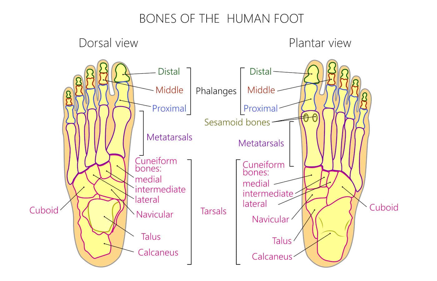
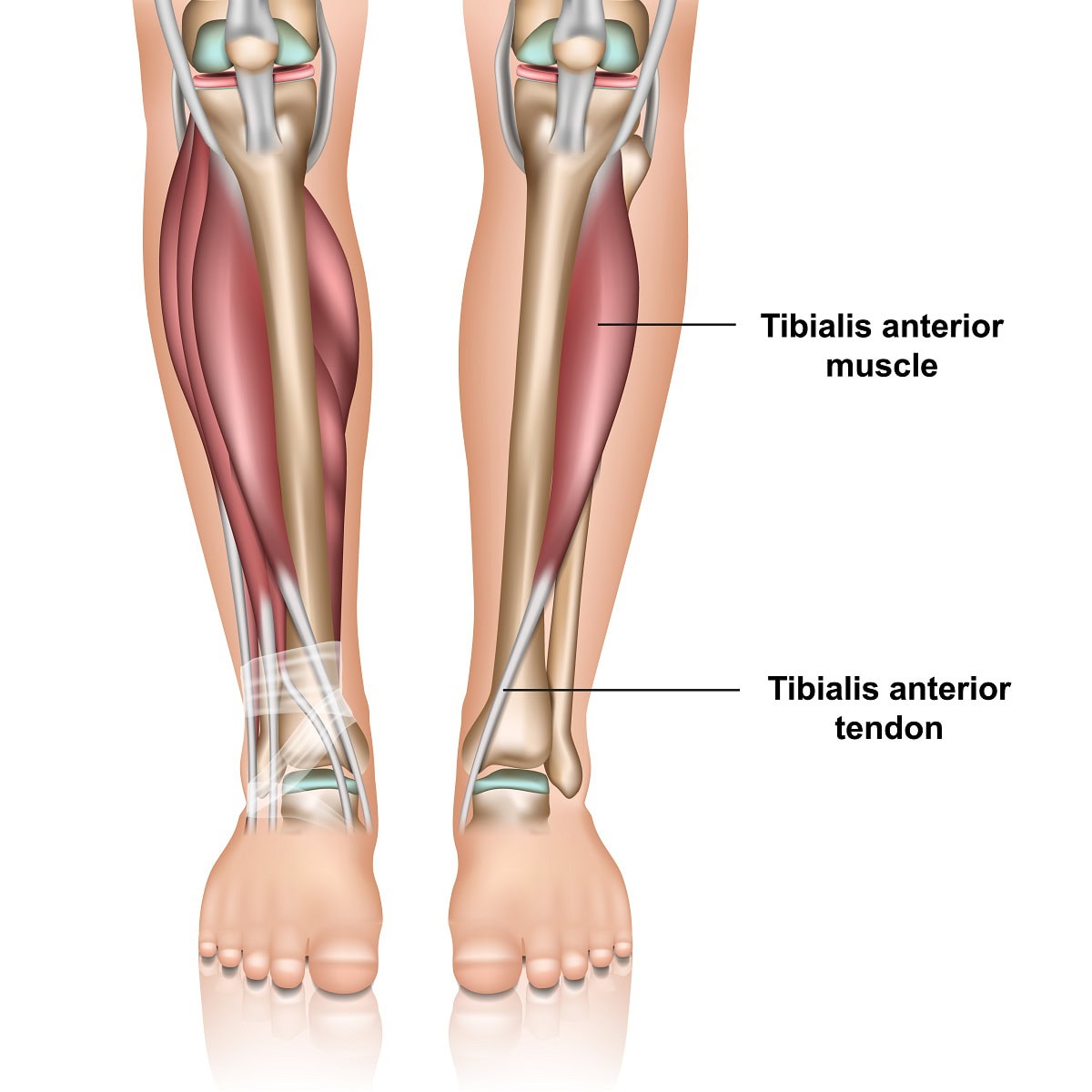
Anatomy and function- The anterior tibial tendon is the third largest tendon in the foot and ankle. The largest is the Achilles tendon and the second largest is the posterior tibial tendon. The anterior tibial tendon originates as a muscle in the lower leg adjacent to the shin, tibia, and extends down to the foot. It is on the front of the ankle. It inserts onto the top and inner side of the arch.

Its function is to pull the foot up and support the arch. It also assists in supinating the foot. Supination is a motion often referred to and means that the arch of the foot increases and the foot angles towards the opposite foot. Finally, it decelerates the downward motion of your foot towards the ground. From the time your heel contacts the ground to the point of the ball of the foot bearing weight it prevents your foot from slapping against the ground. So, it’s very important for normal gait to occur. It helps to identify the tendon by pulling your foot up towards you and it will be the tendon that protrudes on the front of the ankle and is running towards the inside of the foot towards the big toe.

Tibialis anterior tendonitis is a condition that causes pain and discomfort along the front of the lower leg and ankle due to inflammation or degeneration of the tibialis anterior tendon. This tendon plays a crucial role in foot movement, particularly in lifting the foot upward while walking or running.

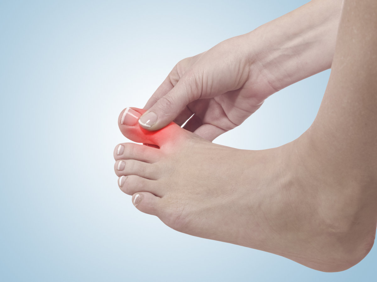
Where Will The Pain Be If I Have Tibialis Tendonitis?

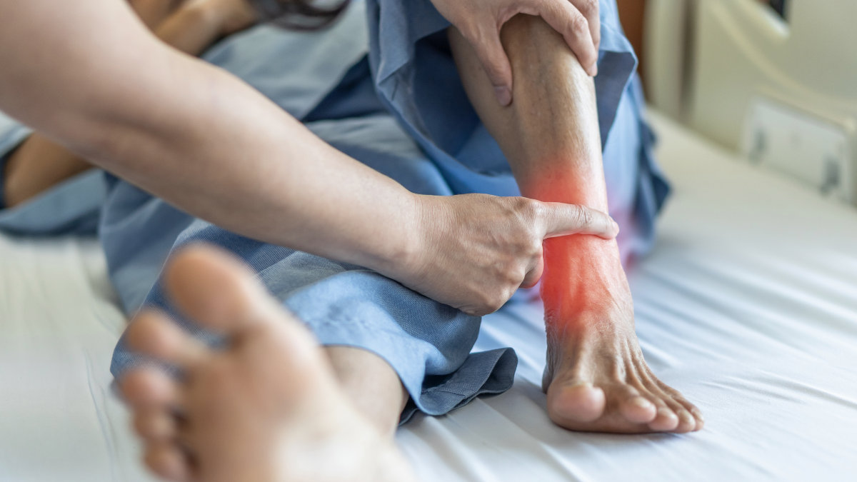
It is most common towards the front of the ankle area. The symptoms of tibialis anterior tendonitis may also create pain above the ankle in the lower leg adjacent to the tibia. This may be referred to as anterior shin splints. Referring to shin splints on the front of the leg. Symptoms may also be closer to or at the point where the tendon attaches to the foot on the inner side of the arch.

What Causes Tibialis Anterior Tendonitis?

Several factors can contribute to tibialis anterior tendonitis, including:

  • Overuse – this may occur from doing too much too soon which can happen in any activity. However, any activity that requires you to extend your foot in front of the knee joint may increase the probability of overuse. Examples of this may be racquet sports, hiking, or running downhill.
  • Biomechanics – If your born with a flat foot the tendon may have to work harder to help support your arch.
  • Improper running technique – Over striding while running can also cause overuse of the tendon. By taking shorter strides you won’t be extending the foot in front of you as much as this will help to reduce the need for the anterior tibial tendon to stop foot slap.
  • Neurological problems – Neurological problems may not cause pain along the tendon, however, may cause damage to the nerves that stimulate the anterior muscle to contract. This is referred to as drop foot. Drop foot can be associated with back issues of a nerve issue around the see. Nerve surgery called decompression surgery can help with this.
  • Trauma – a severe ankle sprain could cause a tear to the tendon. Also, a laceration to the ankle area may also cause a partial or full tear.
  • Shoe gear – High heels may also be a cause and they will put more demand on the tendon to reduce the potential for foot slap because of the elevation of the heel off the ground.
  • Improper Footwear: Wearing unsupportive or worn-out shoes can alter foot mechanics, leading to excessive strain.

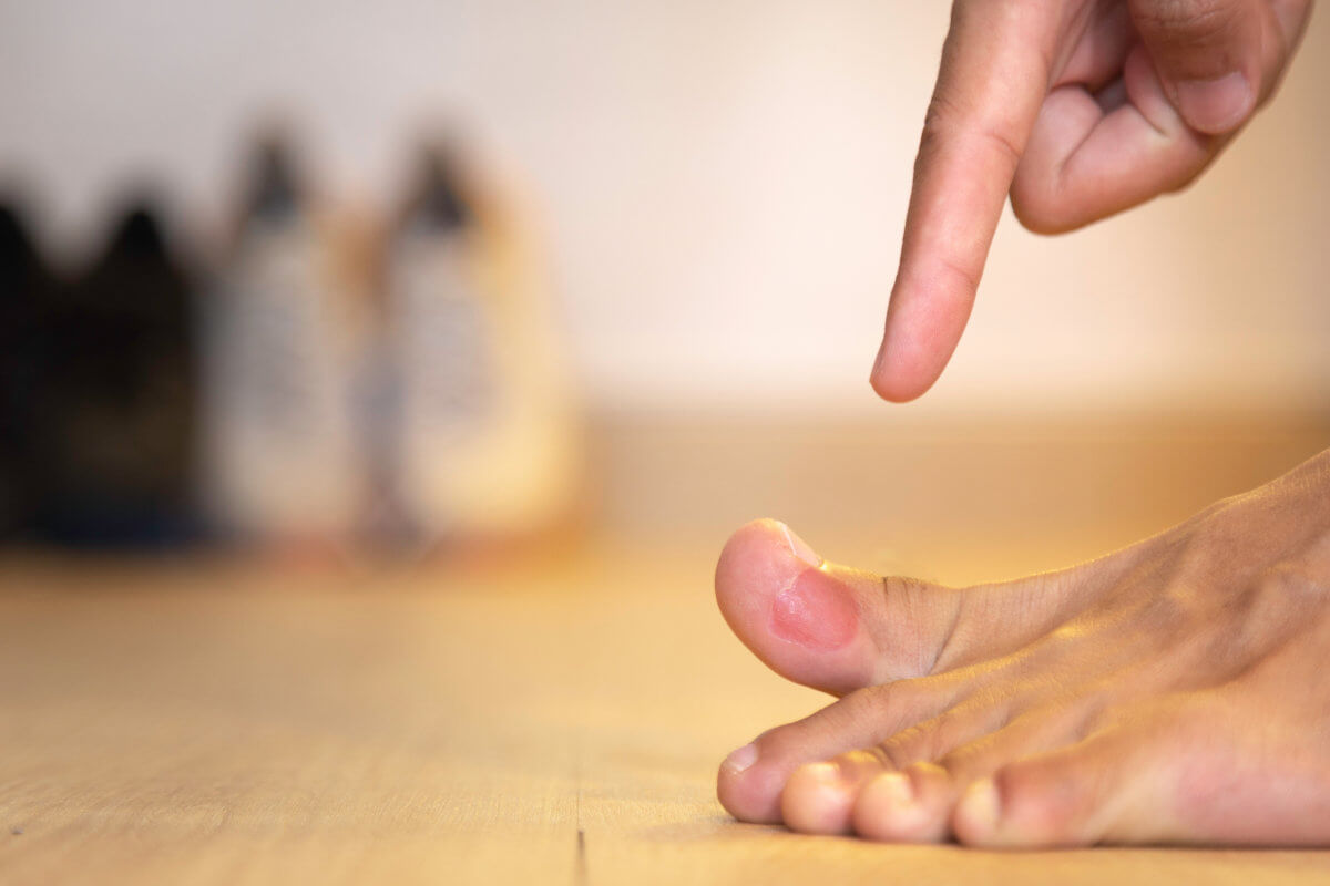
Symptoms of Tibialis Anterior Tendonitis

Common signs and symptoms of tibialis anterior tendonitis include:

  • Pain and tenderness along the front of the shin, ankle, or top of the foot.
  • Swelling in the affected area, especially near the tendon insertion.
  • Weakness or difficulty lifting the foot, potentially leading to a “foot slap” while walking.
  • Stiffness and discomfort that worsens with activity, particularly when walking downhill or running.
  • Pain when pressing on the tendon or flexing the foot upward against resistance.

Diagnosis or Evaluation of Tibialis Anterior Tendonitis

A podiatrist will typically diagnose tibialis anterior tendonitis through:

  • Clinical Examination: The physical examination alone will give the doctor a good idea if the tendon is damaged. There will be pain and swelling along the tendon or its muscle higher up the leg. Also, weakness of the tendon may be noted.
  • X-rays – these will not show tendon damage but are useful to rule out other problems.
  • Diagnostic Ultrasound: Assessing the tendon’s structure and detecting any tears or degeneration.
  • MRI Scan: MRI scans or diagnostic ultrasound are useful to confirm the diagnosis and see the extent of the tendon damage. The diagnosis will frequently be tibialis anterior tendinopathy. The tendon may be damaged but not torn.
  • Gait Analysis: Observing walking patterns to detect abnormalities such as foot drop or imbalance.

Treatments for Tibialis Anterior Tendonitis

Most cases of tibialis anterior tendonitis can be managed with non-surgical treatments, including:

  • Rest and Activity Modification: Reducing strain by limiting high-impact activities.
  • Cold Therapy: Applying ice packs to reduce inflammation and pain.
  • Anti-inflammatory Medications: Nonsteroidal anti-inflammatory drugs (NSAIDs) may help relieve symptoms.
  • Immobilization: Wearing a CAM walker or brace to reduce movement and promote healing.
  • Orthotic Devices: Custom arch supports to help alleviate stress on the tendon.
  • Physical Therapy: Stretching, strengthening exercises, and modalities like ultrasound therapy can aid recovery.

Advanced and Surgical Treatments

For severe cases where conservative measures are ineffective, the following treatments may be recommended:

  • Laser Therapy: Multiwave laser (MLS laser) MLS treatments have shown success in reducing pain and inflammation.
  • Human Cellular Tissue Products: Also known as stem cell therapy, these injections can promote tendon healing. This treatment has significantly reduced the chance of surgery when partial tears are noted. It is important to note that in many cases the tendon may not be inflamed as much as it is breaking down. This is sometimes referred to as tendinosis rather than tendonitis. For this reason, regenerative medicine is very appropriate as it helps to repair the tendon.
  • Corticosteroid Injections (With Caution): While cortisone injections may provide short-term relief, excessive use can weaken the tendon and increase the risk of rupture.
  • Surgical Repair: If a partial or full tear is noted surgery to repair the damage may be recommended. With a full tear weeks of non-weight bearing may be needed. In cases of complete tendon rupture or severe degeneration, surgical intervention may be necessary to restore function. Post-surgery, a period of non-weight-bearing followed by rehabilitation is typically required.

How to Avoid Tibialis Anterior Tendonitis?

It’s important when taking up a new activity to not do too much too soon. Also, if you suffer from flat feet, make sure you have supportive shoes or consider over-the-counter arch supports or custom-made orthotics.

Hopefully this blog will help you to better understand if you have tibialis anterior tendonitis and how some of the newer treatments available including regenerative medicine and laser treatments can help manage the condition. Our foot and ankle clinics cater to patients from Colorado, Wyoming and Nebraska. We also see patients from all over the United States and even our neighbors from Canada who have searched for a ‘expert podiatrist near me‘. Watch our patient testimonials on YouTube as we now have over +11000 followers.

For expert evaluation and treatment, schedule an appointment with Anderson Podiatry Center today.

Call us today at our Fort Collins location (970) 484-4620, Broomfield location (720) 259-5053 or use our online scheduling system to book your appointment.

Hallux Rigidus Surgery: Is a Big Toe Cheilectomy Right for You?

cheilectomy surgery

Cheilectomy is a surgical procedure performed on the great toe to remove spurring from the top of the joint. However, to truly understand cheilectomy surgery we must start at the beginning by explaining a foot problem referred to as hallux rigidus. The surgery is performed to help with pain in the great toe joint and also on top of the great toe joint.

What is hallux rigidus?

Hallux refers to the big toe and rigidus means that the great toe joint is stiff. The great toe is referred to as the first metatarsophalangeal (MTP) joint. Normally it should be able to move 60 to 70 degrees off the ground. With hallux rigidus range of motion of the great toe is reduced. Sometimes it may be 30 degrees and other times motion may be reduced a great deal more, maybe only 10 degrees. Generally, with more restriction of motion there is more pain associated with the problem. In laymen terms what is really occurring is that the great toe joint is becoming arthritic, commonly referred to as osteoarthritis.

Another name that was used to describe hallux rigidus is hallux limitus. It is used to describe the same thing, however the name in recent years has been changed and many doctors agree that hallux rigidus is more severe than hallux limitus. With hallux rigidus you will get bone spurs on the top of your big toe. You may find that your shoes rub this area, and it becomes painful to wear certain shoes. You may also get pain in other areas of the foot, leg, knee or back from favoring the great toe joint. Understand the great toe joint is one of the most important joints of the foot to allow you to walk or run normally.

How does hallux rigidus affect your gait or walk?

When you have lack of motion in the great toe you’ll roll to the outside of your foot as you push off the ball of your foot. You have to do this because the joint motion is limited so you have to get motion elsewhere and you achieve this by rolling onto the ball of the foot by the smaller toes. The modification in your gait may cause all kinds of other aches and pains. Frequently the first think to happen is you start to notice pain in the great toe, but you may adapt the pain by favoring the great toe thereby reducing the pain in the toe joint and increasing pain in other areas up into your ankle knee or hip joint. But it all started in your dysfunctional great toe.

What causes hallux rigidus?

  • Trauma – you may have had a severe sprain or jamming of the great toe joint. You may also have had a fracture in the joint. Both can damage the cartilage in the joint and therefore start the arthritic process.
  • Other arthritic conditions – If you have chronic gout your gout attacks frequently occur in the great toe joint. Because a gout attack causes severe inflammation in the great toe joint this will cause damage to the cartilage. The same idea holds true with autoimmune conditions such as rheumatoid arthritis.
  • Improper biomechanics – This is by far the most common cause. For the big toe to move up to 60-70 degrees of the ground two motions need to occur. First the great toe, hallux will move up 20 degrees. However, the first metatarsal bone forms the portion of the great toe joint that you bear weight on needs to push down against the ground. When the first metatarsal head pushes down against the ground the great can now glide up higher another 40 degrees to get up to 60-70 degrees of motion. It’s as if there are two axis of motion. One axis of motion allows the toe to move up 20 degrees then when the first metatarsal bone moves downward the great toe can move upwards even more. If the first metatarsal cannot move down then the great toe will jam as it tries to move upward. This jamming on the top of the joint is what causes spur formation and also wearing down of the cartilage on the upper portion of the joint. So now you may have pain from the spurring on the top of the joint rubbing shoe gear, and pain on motion of the joint as it jams as it moves upwards.

Non-surgical treatments to avoid cheilectomy surgery

  • Anti-inflammatory – To reduce pain and swelling Anti-inflammatory drugs may be used in the early stages as a pain reliever. However, long term use may be detrimental to the health of the joint.
  • Orthotics – depending on the type of custom-made inserts called orthotics are very helpful in the early stages of Hallux rigidus as the help to combat the biomechanics the promote the condition and may offer some pain relief. If the type of orthotic you’re using is store bought or not created from a non-weight bearing impression of your foot it may not be effective.
  • Regenerative medicine – The use of human cellular tissue products may be used to successfully eliminate pain in the joint and delay or possibly eliminate the need for surgery in some cases. Many of you are aware that stem cell treatments are commonly done for arthritis knees however very few realize this is an option for the great toe joint. See below to understand why the great toe has more opportunity to repair itself. And please watch this video.

What is involved in cheilectomy surgery?

Cheilectomy surgery is a procedure involving removal of the excess bone on the top of the joint and revising the joint of the big toe. If there is significant damage to the cartilage in the joint, then microfracture technique may be part of what the surgeon does during surgery. By making small drill holes(microfracture) in areas of the joint where cartilage is worn down to bone this will allow blood into the joint after surgery and in doing so allows repair of the damaged cartilage. The cheilectomy surgery technique works very well on the great toe joint. It is believed that cartilage has a difficult time repairing when there is vertical load on the cartilage. This would be the situation for a knee or hip joint. However, there is very little vertical load on the great toe joint the microfracture technique will have more potential benefit.

Post operative recovery – most patients are able to walk immediately after surgery in a surgical shoe or Cam walker. Sutures are usually removed between 2-3 weeks after surgery so the foot will have a dressing on during that time and the foot will need to be kept dry. Most patients are going back to shoes in 3 weeks and returning to some activities by 4 weeks and more full activities by 6-8 weeks. The risk of this surgery are not as significant as other foot surgery but you should be aware of them as problems such as blood clotting referred to as deep vein thrombosis may occur.

How to reduce the chances of joint fusion or joint replacement?

It needs to be stressed that this procedure may not be the end of your great toe pain. It’s possible for the cheilectomy procedure to work for years but the arthritic condition continues. Pain returns as the joint becomes more damaged and needs to be replaced or fused.

Regenerative medicine – The big toe joint has an unfair advantage for stem cell treatments to work. This is because the vertical load on a joint inhibits repair on joint cartilage and the great toe joint has very little vertical load. The procedure is simple to do. It’s done in a clinic setting and you’re able to walk immediately returning to regular shoes in 48 hours with no pain pills needed. Also, it important to consider before you symptoms become severe

Surgical treatment – If surgery is considered earlier, it is better to do a cheilectomy. First you need to have pain to consider a surgical approach. However, removal of the bone spurs and revising the joint with the microfracture technique may have a greater benefit long term if its done before the damage to the joint has become severe.

In summary

Cheilectomy surgery may not be the only surgery option, but if you wait too long it could result in hallux rigidus.

Regenerative medicine has an important role in reducing or delaying the need for surgery including fusion and joint replacement. And should be done early.

Schedule your appointment online or call our Fort Collins and Broomfield, Colorado locations to connect with a trusted foot doctor near you at our renowned foot and ankle clinic.

Frequently asked questions

1) What is a big toe cheilectomy, and how does it help with hallux rigidus?

A big toe cheilectomy is a surgical procedure that removes bone spurs and part of the arthritic joint to improve movement and reduce pain in patients with hallux rigidus—a condition that causes stiffness and pain in the big toe. It is most effective in the early to mid-stages of the condition, helping to preserve joint function and delay the need for more extensive surgery.

2) Who is a good candidate for a big toe cheilectomy?

This procedure is ideal for patients with mild to moderate hallux rigidus who experience pain and restricted motion due to bone spurs but still have some cartilage remaining in the joint. If arthritis is severe or joint damage is extensive, alternative procedures like joint fusion (arthrodesis) or joint replacement may be more suitable.

3) What is the recovery time after a cheilectomy?

Recovery typically takes 6 to 8 weeks. Most patients can walk with a stiff-soled shoe within a few days but should avoid strenuous activities for several weeks. Swelling and stiffness may persist for a few months, and physical therapy may be recommended to restore mobility and strength.

4) What are the risks and potential complications of this surgery?

As with any surgery, risks include infection, nerve damage, prolonged swelling, or incomplete pain relief. In some cases, arthritis may continue to progress, requiring future procedures. However, most patients experience significant pain relief and improved mobility after surgery.

5) Will I be able to return to sports and physical activities after a cheilectomy?

Many patients can resume low-impact activities such as walking and swimming within a few weeks. However, high-impact sports like running and jumping may take longer and could be limited if arthritis worsens. Your surgeon will guide you based on your healing progress and activity goals.

Call us today at our Fort Collins location (970) 484-4620, Broomfield location (720) 259-5053 or use our online scheduling system to book your appointment.

Conquer Chronic Ankle Instability: Your Guide to Stronger Steps!

chronic ankle instability

Ankle sprains are a common foot and ankle injury. When ankle sprains recur frequently or if not treated properly this can lead to chronic ankle instability. Proper treatment and evaluation are important as ankle sprains tend to be undertreated and not always thoroughly evaluated.

Basic anatomy of your ankle

The outside of the ankle is supported by three ligaments that connect the smaller leg bone, the fibula, to the bones of the foot.

Most frequently damaged ligaments

The anterior Talofibular ligament ATFL – The connects the fibula to the ankle bone called the talus. It runs forward from the ankle parallel to the ground. It’s function is to keep your foot from moving forward out of the ankle joint

Second most frequently injured

The calcaneal fibular ligament CFI- This connects the fibula to the heel bone and travels vertical to the walking surface. This ligament keeps your foot from rolling out of the ankle joint. It supports the subtalar joint which is the joint beneath the ankle. Its function is like a universal joint on a car. It allows your foot to move in different body planes. When you make big circles with you foot most of that motion is coming from the subtalar joint.

The posterior talofibular ligament – This ankle ligament connects the fibula to the talus the ankle bone and runs towards the Achilles tendon. This ligament is the least frequently injured.

Why do ankle sprains occur?

  • Activities – certain risk activities are more likely to put you at risk of an ankle sprain. Any sports that require side to side motion such as racquet sports, tennis and pickleball. Sports that are ballistic and involve contact such as basketball, football and soccer. Activities that involve hiking over irregular terrain can also be riskier.
  • Shoe gear – shoes that are broken down and not as supportive may also lead to a sprain.
  • Foot types – Certain foot biomechanics are more likely to lead to chronic ankle sprains.
  • Ligamentous laxity – some are born with ligaments that are very flexible such as Erlos Danlos syndrome.
  • Neurological issues – you may have a mild case of drop foot from nerve damage. This could be from common peroneal nerve entrapment. The muscles that pull your foot upwards and sideways are weaker because of this condition.

Evaluation for chronic ankle instability

  • Proper evaluation starts with a good history – The mechanism of your injury will likely help the doctor to determine what structures may be injured.
  • Physical examination – There will usually be pain and swelling over the ligaments that are frequently injured.
  • X-rays – these may be taken to insure there are no fractures.
  • MRI – If a severe sprain takes place then imaging studies will be useful to determine the severity of the ligamentous damage.

Chronic ankle instability Treatments

This may require an extended period of time to rest the injury.

  • Immobilization– depending on  the severity of the injury the doctor may recommend the use of a Cam walker to allow the damaged ligament that were torn to repair properly.
  • Physical therapy– to help promote healing and strengthen the muscles around the ankle. This can also strengthen the peroneal tendons that support the outside of the ankle.
  • Ankle bracing– this may be used during healing and even for a longer period of time to protect the area from another sprain as you return to full activity
  • Anti-inflammatories– these may help with pain and swelling during the early stage of the ankle sprain.
  • Orthotics– custom made arch supports can help provide a stable base for the foot to stand on in the shoegear and reduce the frequency of recurrent ankle sprains.
  • Surgery-Surgery may be performed to repair the ligaments that have become damaged and weakened and no longer provide support to the ankle. The ligaments may be repaired along with using tendons around the ankle to increase support to this area. This can prevent repeated ankle sprains.
  • Nerve surgery– if it is from weakness because of neurological issues decompression of the common peroneal nerve may be performed.  The common peroneal nerve tunnel can become tight, or the nerve tunnel could be damaged. Reference for my research paper.

What will happen if left untreated?

With repeated ankle sprains the possibility of getting arthritis in the ankle or subtalar joint beneath the ankle joint increases. Treatment for the ankle arthritis may include ankle scope surgery to clean out the damage in the ankle, Stem cell treatment with human cellular tissue products, see video below.

Two suggestions to help prevent the chance of having chronic ankle instability.

Proper evaluation and treatment

The first and most severe sprain you have is the most important to properly treat. A common mistake I see is patients may not get an ankle sprain looked at by a doctor. Or if they do they may go to the emergency room or urgent care. X-rays shoe there is not fracture. They may leave with an ace wrap or ankle brace and are told to ice it. In other words ankle sprains are under treated and under evaluated.

Ideally if it a severe sprain with a good history taken by the doctor and a complete exam and in some cases and MRI a better treatment plan could be determined. If it’s a third degree sprain meaning one or more ligaments of the ankle are fully ruptured than immobilization of the ankle may allow the ligaments to fully repair and retain there strength. So when in doubt see a foot and ankle specialist.

Consider trail running or hiking

Yes, I know this may sound contradictory and I’m speaking from my own experience. I’ve run all my life from college, through medical school and all the way through my sixties. However, starting in my sixties, I gave up running on concrete and asphalt. I started train running as it really reduced the wear and tear on my body from running on hard surfaces. I was amazed. However I was very afraid at first as I was prone to ankle sprains. I have found that I’ve dramatically reduced the number of sprains I have, and I believe its for three reasons.

  1. I’m extremely efficient and looking where I step every time! I got very lazy doing this on smooth surfaces.
  2. Muscle strength in my lower legs has increased. This is simply because the muscles that have to support my foot from moving side to side are being used more and I believe it protects me from having sprains and I can make a faster correction when I sense I’m about to sprain my ankle.
  3. I have found that trail running shoes are important to use. They are treads that have more grip and have lower profile which I believe lowers the center of gravity of my foot in the shoe. It also allows me to feel irregularities in the terrain and make adjustments better.

So remember to get a professional certified local foot doctor’s opinion when in doubt and consider the suggestions for ankle sprain treatment and prevention in this blog. If you live in Colorado or nearby states consider making at appointment at our Broomfield or Fort Collins foot clinic. Our staff is compassionate and caring towards all our patients. We also have several testimonials and have now surpassed 5000 subscribers on our Anderson Podiatry Youtube channel.

Call us today at our Fort Collins location (970) 484-4620, Broomfield location (720) 259-5053 or use our online scheduling system to book your appointment.

What is Fibularis Longus Pain and Why Proper Evaluation is Needed?

fibularis longus pain

Anatomy -The fibularis longus muscle is one of two muscles that help support the ankle joint on the outside of the ankle. The other muscle is fibularis brevis. The fibularis longus and brevis may also be referred to as the peroneus longus and the peroneus brevis. Both of the peroneal muscles originate on the outside of the leg and extend down towards the ankle. At the level of the ankle joint they become tendons and act like stirrups around your ankle. Their function is to stabilize your ankle from moving side to side. The fibularis longus also helps to support the arches of the foot. When you activate these muscles, the foot will be pulled away from the opposite foot. When you have pain of the fibularis longus it is usually the tendon around the ankle and foot rather then the muscle portion that extends up into the lower leg.

Tendonitis versus tendinosis versus tendon tear.

When evaluated for this problem it’s important to establish which one of these conditions you have

  • Tendonitis – this simple means the tendon is inflamed and occurs early in the process.
  • Tendinosis -This means that the tendon is damaged and may have small tears within the tendon.
  • Tear – This is the most advanced presentation. There is an obvious tear and usually extends along the length of the tendon.

Where is the pain located – Both the fibularis longus and brevis run along the outside of the ankle posterior to the lateral malleolus. See diagram below.

peroneus muscle brevis fibularis longus inforgraphic

When there is pain it will be located along the outside of your foot and ankle by your heel bone and may extend up towards the outside of the ankle just behind the fibula. There will be pain and swelling along the length of the tendon.

What is the cause of fibularis longus pain?

  • Overuse – simply doing too much too soon.
  • Activity on uneven surfaces – because these tendons support the ankle from moving side to side walking on irregular terrain will overuse the tendons. Also, activities like tennis, pickleball and skiing require side to side motion and are more likely to stress the tendons.
  • Trauma – when a ankle sprain may cause peroneal tendon injuries. As most ankle sprains involve rolling onto the outside of the foot injury may occur. The severe forces generated can cause pain to persist in this area even after the pain from the ankle sprain no longer exists.
  • Foot types – certain foot types may make you more prone to overuse of the tendons. This could include severe flat feet but also very high arched feet.
  • Shoe gear – you may be wearing shoes that are worn down or do not offer enough support.

How do we evaluate and diagnosis?

  • History – getting a good history from the patient is important. Was there an injury, how long have you had the problem, have you recently changed your activities. What have you tried.
  • Location of the pain – The physical examination is very important. Usually there will be pain and swelling along the tendon.
  • Diagnostic ultrasound – we can evaluate the tendon by using ultrasound to look for tears or inflammation along the tendon
  • X-rays – They may be used to rule out bone problems that could mimic or be associated with pain.
  • MRI – This is very useful to confirm whether there is a tear and its severity.

Treatments for fibularis longus pain

  • Rest and immobilization – in acute situations where the pain is very recent and there is no apparent tear using a Cam walker to minimize motion in the area can help.
  • Anti inflammatory medications – These may be helpful especially if in the acute stages.
  • Orthotics – custom made inserts can be very helpful as they will help support the foot from moving side o side and therefore eliminate the pain.
  • Physical therapy – using modalities such as ultrasound may also help Stretching and mobilization exercises may also be used.
  • Laser treatment – We use a very effective laser called MLS laser which is also used as part of a treatment plan.
  • Surgery – if there is a tear and conservative treatments fail this can be very successful. It will require lay up of at least four weeks of immobilization.
  • Regenerative medicine – using stem cell treatments has been very successful and has greatly reduced, almost eliminated the need for surgery for many of our patients. This will aid in reducing inflammation and repairing the tendon. Patients are in Cam walker for four weeks and can walk.

The dangers of not knowing if you have a tear

The most important thing to understand is that the majority of fibularis longus pain may not be from tendonitis with tendon inflammation but rather tendinosis. Tendons tend to have less blood supply than other structures. So when they become over stressed microtears may occur. So its very important to know if this is taking place. How do we know if the tendons inflamed or breaking down. MRI is the answer.

The dangers of physcial therapy without proper evaluation

How does treating a tear differ from tendonitis. If the patient has a tear that physical therapy with the potential for stretching would not be advisable. This could further injure the tendon. I’ve seen this in practice. Patients are going to physical therapy and peroneal tendonitis was diagnosed because of pain alone the tendon. The physcial therapist may recommend stretching and putting the ankle area and the tendon through ranges of motion that could further damage the tendon. The problem is made worse. If they had known there was a tear the option of regenerative medicine would be considered, and the possibility of surgery reduced.

What is the Recovery Time for Peroneal Tendonitis?

The recovery time for peroneal tendonitis varies depending on the severity of the condition and the effectiveness of the treatment. Generally, with proper rest, physical therapy, and treatment, mild to moderate cases of peroneal tendonitis may take about 4 to 6 weeks to heal.

For more severe cases, or if there has been chronic damage to the tendons, recovery could take several months, potentially 3 to 6 months, especially if surgery is required. Early diagnosis and adherence to a treatment plan, including rest, ice, compression, elevation (RICE), physical therapy, and possibly orthotics or bracing, can help speed up the recovery process.

What are some Exercises one can do if they have Peroneal Tendonitis?

If you have peroneal tendonitis, gentle exercises can help strengthen the tendons and support recovery. Here are some exercises that are commonly recommended:

  1. Ankle Circles
    • How to Do It: Sit with your leg extended and slowly rotate your ankle in a circular motion. Make sure to go in both clockwise and counterclockwise directions.
    • Repetitions: 10-15 circles in each direction.
    • Benefit: Improves range of motion and reduces stiffness.
  2. Towel Stretch
    • How to Do It: Sit with your leg extended, loop a towel around the ball of your foot, and gently pull the towel towards you, keeping your leg straight.
    • Hold: 15-30 seconds.
    • Repetitions: 2-3 times.
    • Benefit: Stretches the calf muscles and Achilles tendon, which can help reduce strain on the peroneal tendons.
  3. Calf Raises
    • How to Do It: Stand with your feet shoulder-width apart and slowly rise up onto the balls of your feet, then lower back down.
    • Repetitions: 10-15 repetitions, 2-3 sets.
    • Benefit: Strengthens the calf muscles, which support the peroneal tendons.
  4. Resistance Band Eversion
    • How to Do It: Sit down and secure a resistance band around your foot. Hold the other end with your hand or attach it to a stable object. Slowly move your foot outward against the resistance of the band.
    • Repetitions: 10-15 repetitions, 2-3 sets.
    • Benefit: Strengthens the peroneal muscles, which helps stabilize the ankle.
  5. Heel-to-Toe Walking
    • How to Do It: Walk in a straight line by placing the heel of one foot directly in front of the toes of the other foot. Keep your balance and move slowly.
    • Distance: Walk 10-15 feet, 2-3 times.
    • Benefit: Improves balance and strengthens the muscles around the ankle.
  6. Balance Exercises
    1. How to Do It: Stand on one leg with your eyes open and hold for 30 seconds. As you progress, try closing your eyes or standing on an unstable surface like a cushion.
    2. Repetitions: 2-3 times on each leg.
    3. Benefit: Enhances ankle stability and proprioception.
  7. Alphabet Exercise
    • How to Do It: While sitting, lift your foot off the ground and try to “draw” the letters of the alphabet in the air with your toes.
    • Benefit: Increases ankle mobility and reduces stiffness.

Note: Before starting any exercise program, it’s important to consult with your physical therapist to ensure the exercises are appropriate for your specific condition and won’t exacerbate your symptoms. Start slowly, and if you experience any pain during these exercises, stop immediately and consult a podiatrist near you.

So if you’re having pain on the outside of your ankle or foot consider fibularis longus pain as a possibility. Early evaluation and treatment for peroneal tendonitis is needed. Proper evaluation is also important as a tear (tendinosis) versus inflammation (tendonitis) are treated differently. Make sure you consider seeing a foot specialist and consider getting an MRI if you’re not getting better.

Give us a call to talk to your certified and experience local foot doctor in Fort Collins and Broomfield, Colorado.

Diagnosis and Treatment of Freiberg’s Disease: Pain in the Ball of the Foot

Freibergs disease second metatarsal head

Freiberg’s disease is a rare presentation in the foot that can be misdiagnosed and overlooked. To better understand what it is we first need to go over the basics of how your bones develop in your foot.

What are the symptoms of Freiberg’s disease?

The patient will have pain in the ball of the first. The area that is affected is the metatarsal head. Pain is most common at the base of the second metatarsal head. and rarely at the 4th or 5th toe. There may be swelling at this joint. There may also be a limited range of motion compared to the adjacent joint. Rolling up on the ball of the foot when walking may create pain in this area.

weight bearing bones phalanges freiberg's disease second matatarsal head

The basics of growth plates in the foot

You may have heard of a term called growth plates. We all have them as our bones develop in childhood. A growth plate is in the long bones of our body and it’s from this region the bone develops to make a short bone longer. The growth plate is comprised of cartilage. If you look at an x-ray of a child’s foot you see a dark area going across the bone which contrasts with the white appearance of the bone. Cartilage does not show up on x-ray and that explains the dark area. For a bone to get longer the cartilage cells in the growth plate area multiply and then transform into bone cells and over time the bone will elongate to its adult length. Then at puberty the growth plates will close, meaning the cartilage that was once there will transform into bone. The growth plate will no longer be seen on an x-ray.

What causes Freiberg’s disease?

When Frieberg’s disease occurs the growth plate has been damaged. With this damage the blood supply to the growth plate may occur. This will then create what is referred to as avascular necrosis to the metatarsal head which is the area where the growth plate is located. The metatarsal bones are the long bones that goes from the midfoot area down to the ball of the foot. Metatarsal heads are the bones you bear weight on in the ball of the foot. The national library of medicine has labelled it as “an uncommon yet clinically significant condition“.

As the word implies, blood supply is diminished (avascular) and the bone will die away (necrosis). With diminished bone directly beneath the cartilage of the metatarsal head the cartilage deteriorates. This cartilage damage will cause them to become arthritic. There are two primary reasons this may happen.

  1. Trauma to the area in childhood. The story that I will commonly hear is that I got stepped on, many times by a horse, but any type of direct blow to this area could damage the growth plate that then goes undetected.
  2. Overload on the metatarsal head – if the metatarsal is longer in relation to the neighboring metatarsals the stress in this area as the bone is developing can also be a cause. This is especially true in the second metatarsal. It may also be from wearing high heels that would overload the second metatarsal.

What is done to diagnose the problem of Frieberg’s disease?

  • clinically there will be pain at the metatarsal phangeal joint involved. The motion may be limited and painful to move.
  • X-rays will show evidence that the joint space may be narrower, and the metatarsal head may appear flatter than normal.
  • MRI- An MRI can more specifically evaluate the joint and reduction of blood supply to the area.

Treatments for Freiberg’s disease

Conservative treatment

  • Immobilization – resting the area by using a Cam walker for 4-6 weeks (about 1 and a half months) may be helpful. However, this is unlikely to give long-term relief.
  • Orthotics – The use of custom-made inserts may help to reduce the load on the joint and thereby reduce symptoms
  • Stiff sole shoes – although it may be impractical, shoes that don’t flex in the ball of the foot can also give relief. Avoid high heeled shoes-These will place more weight on the ball of the foot placing stress on the metatarsal head.
  • Regenerative medicine – As the joint is arthritic use of human cellular tissue products such as placenta cell and umbilical cord may also be considered.

Surgical treatments for Freiberg’s disease

The following surgical approaches will depend on the severity and age of the patient

  • Arthrotomy- This means the joint is opened and the damaged cartilage is removed. There may also be pieces of cartilage that have broken off that are floating in the joint. These are called loose bodies. In the area where there is no cartilage small drill holes can be made, and this will help promote potential new cartilage formation after surgery. This is referred to as the microfracture technique.
  • Removal of the joint – by removal of metatarsal head or a portion of the phalanx bone pain can be relieved.
  • Osteotomy – after the damaged cartilage is removed a bone cut is made in the metatarsal to reposition it so it can now articulate in an area of the joint that has better cartilage.

So, if you’re struggling with pain in the ball of your foot and are already seeing a doctor, consider that you may have Freiberg’s disease. Of all the foot ankle issues Freiberg’s disease is not a common problem so many doctors may not be looking for it or doing the proper evaluation to diagnose it.

Call us today at our Fort Collins location (970) 484-4620, Broomfield location (720) 259-5053 or use our online scheduling system to book your appointment.

Phalanges of the Foot: A Guide to Their Complex Anatomy

phalanges in the foot

There are many bones in the foot making its anatomy one of the more complex in your body. Of the 26 bones in the human foot approximately half of them are phalanges in the foot. Generally, as we start towards your ankle joint and move down the foot towards the toes the number of bones become more multiple in the distal extremity. For example, in your rearfoot there are two large bones the talus (ankle bone) and calcaneus (heel bone) and 14 phalanx bones. The anatomy of the hands and feet are similar but because you walk on your feet one could argue they are more of a structural wonder. We will describe the anatomy of these bones and potential issues that could cause pain in this area of your foot.

Basic anatomy of phalanges in the foot

The phalanges are the bones of the foot that comprise the bone of your toes. They are connected to the larger and longer metatarsal bones that extend from the midfoot down to the ground forming the ball of your foot. When you bear weight on the ball of the foot you’re standing on the metatarsal heads on the plantar surface of these long bones. The phalanges extend beyond the ball of the foot forming the digits. They are comprised of the proximal, middle and distal phalanges.

Proximal phalanges of the feet

These bones form the articulation with the metatarsal bone These two bones form the metatarsal phalangeal joints. These five joints that form the ball of your foot have a lot of upward mobility called dorsiflexion. This allows these joints to bend upwards to allow you to pivot up onto the ball of the foot when you walk or run. The anatomy of these joints is very complex as they require various tendons and ligaments to maintain their stability. This is especially true for the great toe joint. This has beneath it two bones called sesamoid bones that glide beneath the great toe joint as the toe moves up and down, much like your kneecap. The function to improve leverage and therefore strength to the great toe joint as this joint is important to assist in propelling us forward when walking or upward when we jump.

Intermediate phalanges

The next phalangeal bones as we move away from the ball of the foot are the intermediate phalanges. They may also be called the middle phalanges as they lie in the middle of the toes. They are smaller than the proximal phalanges bones. As was just mentioned the great was unique as it has two bones beneath it called the sesamoid bones and there is one more difference with the great toe. The great toe only has two phalanx bones. They are referred to as the proximal and distal phalanx bones.

Distal phalanges

At the very end of each toe are the distal phalanges. These are the smallest of the phalanges and lie beneath the toenails. Rather than having a tubular shape like the proximal and intermediate phalanges they are short and triangular shaped.

The joints of the toes.

We had mentioned earlier that the joint formed by the metatarsal bones and the proximal phalanges is called the metatarsal phalangeal joint.

  • The joint formed by the proximal and intermediate phalanx is called the proximal interphalangeal joint (PIPJ).
  • The joints formed by the intermediate phalanx and the distal phalanx are called the distal interphalangeal joints (DIPJ).
  • Finally, the great toe must be different as it only has two phalanges, so it does not have a proximal and distal interphalangeal joint. Because it has only two phalanges its joint between these two bones is called the interphalangeal joint.
  • Finally, the great toe also has its name, referred to as the Hallux.

To summarize the big toes are different in three ways

  1. It has only two phalanx bones.
  2. It has two sesamoid bones beneath it at the metatarsal phalangeal joint.
  3. It has its own name, Hallux.

Functions of the phalanx bones in the foot

These appendages help with your balance and push your foot off the ground when you walk, run, or jump. The great toe is more important for these purposes than the other four toes. It’s important to note that in a situation of an amputation of a digit. Removal of one of the smaller toes will have little effect on your balance and ability to function normally. However, the absence of the great toe has more impact.

Problems that can arise in the phalanges

Since these bones are the distal extremities of the foot, they can be subject to trauma such as stubbing your toe. When this happens you may fracture a phalanx bone. Contrary to the popular notion that nothing can be done it’s important to have the injured toe evaluated. Why? If a fracture is present a common treatment will be to buddy tape the injured toe to an adjacent toe. This is important during the healing phase to ensure the fracture does not get too displaced. A fracture that heals in a misaligned position can create long-term pain and require surgical intervention. When the great toe is fractured, proper evaluation is needed to ensure proper healing because of its importance for normal ambulation. Again, proper treatment and follow-up is important.

Hammertoes

Hammertoes are deformities of the digits and can create pain from the dorsal surface of the toe rubbing shoe gear of the walking surface. There are three types of hammertoes
Mallet toe. This deformity is at the distal interphalangeal joint. The metatarsal phalangeal joint and proximal interphalangeal joint are in good alignment; however, the distal interphalangeal joint is flexed downward.

Claw toe. This deformity involves all the joints. The metatarsal phalangeal joint is flexed upwards, and the proximal and distal interphalangeal joints are flexed downward. The bases of the middle phalanx may rub shoe gear.

Hammertoe this deformity involves two joints. The metatarsal phalangeal joint is flexed upwards, and the proximal interphalangeal joint is flexed downward.

Treatment for hammertoes is

  • Conservative – wearing appropriate shoes with bigger toe box, padding, etc.
  • Surgery – Foot Surgery is performed to realign the digit to its normal position.

Hopefully, this blog has given you more insight into the phalanx bones, their anatomy, and problems you may encounter in this region of your foot. If you are seeking a ‘podiatrist near me’ in Broomfield or Fort Collins, consider Anderson Podiatry Center. We have our own surgical foot center that has many advantages which you can review here.

Call us today at our Fort Collins location (970) 484-4620, Broomfield location (720) 259-5053 or use our online scheduling system to book your appointment.

What Is Sesamoiditis And What Is The Best Sesamoiditis Treatment?

sesamoiditis treatment anderson podiatry center

The sesamoid bones are small pea shaped bones that sit beneath the big toe joint. Where there is pain under your big toe it may be called sesamoiditis pain. Let’s look at why you may be seeking sesamoiditis treatment to resolve a common foot pain.

Sesamoiditis basics – The sesamoid bones are like the kneecap, patella bone. They sit beneath the big toe joint and the great toe, and both are attached to muscles and tendons that help pull the big toe downward. Much like the kneecap they each glide in a groove under the joint. They function to increase the leverage of the muscles of the foot to pull this toe downward. This motion is very important for normal gait while running or walking. You may have heard of the term turf toe, this is associated with the sesamoid bones but may also involve inflammation to the tendons and muscles that attach to the sesamoids.

What are the symptoms of sesamoiditis?

Sesamoiditis pain tends to be very localized beneath the big toe joint in the ball of the foot. You’ll favor your foot tending to avoid putting pressure on the bottom of the great toe. High heels will make the pain worse. You may have swelling associated with it. The pain tends to be deep and dull, and you may notice it the most when you first step down in the morning. Which is why most people try to pass it off as morning stiffness and not seek sesamoiditis treatment.

What is the cause of sesamoiditis?

  • Foot type – some feet may tend to roll inward referred to as pronation. This would cause more pain to be placed on the ball of the great toe.
  • Trauma, sesamoid fractures. You may have fractured the sesamoid bone. You’ll usually be aware of this as a lot of force would be needed to cause a fracture. Landing with a lot of weight on the great toe or severely jamming your great toe upwards may cause a fracture.
  • Bunions – A bunion deformity causes the great toe joint to be misaligned. Because of this the seamaids may no longer glide centered in the grooves beneath the metatarsal bone but may now be gliding over centered. This may result in erosion of the cartilage on the ridge that separates the two grooves. Both the cartilage on the seamaid bone and the cartilage on the bottom of the metatarsal breakdown causing octopartite to occur
  • Hallux Limitus – This is a deformity of the great toe joint that limits range of motion of the great toe joint. As the joint starts to reduce its motion this may also interfere with the normal gliding of the sesamoid bones and produce pain.
  • Overuse- This may be the most common cause and typically develops gradually. This is especially true in activities that may cause a lot of weight bearing beneath the big toe such as ballet, dancing, basketball and volleyball to name a few.
  • Shoe gear – If you wear high heels excessively it may cause sesamoiditis. Soft soled shoes would be best.

How is sesamoiditis diagnosed?

  • Physical examination – your podiatrist will evaluate your foot to see if you have pain and swelling where the sesamoid bones are located.
  • x-rays- these will be taken to see if the sesamoid bones appear normal.
  • Congenitally abnormal – you may have been born with an enlarged sesamoid or one that is bipartite, has two parts. These abnormal presentations make it more likely to have pain from these sesamoids.
  • Alignment – are the sesamoids positioned where they should be beneath the great toe joint. A bunion deformity will cause them to be misaligned and more likely to hurt.
  • Fracture – the x-rays will also help to rule out a fracture.

How can I avoid sesamoiditis pain?

  • Surgery – Surgery to remove the pain of damaged sesamoid may also be performed but only after conservative measured have failed. When surgery is performed it is usually the medial sesamoid that tends to be more prone to pain. This sesamoid is the one that is located towards the other foot.
  • Reduce or eliminate activities – for some it may be practical to eliminate an activity that causes more stress to this area of the foot. For others better management of how much of the specific activity that is more likely to cause an injury should be considered. You should gradually increase each activity or consider doing it less frequently.
  • Avoid high heels – these will tend to put more weight on the bottom of the great toe joint.

What is the Best Surgical and Non-Surgical Sesamoiditis Treatment?

Non-Surgical Treatments

  • Rest and Activity Modification – Reducing weight-bearing activities to allow the sesamoid bones to heal.
  • Orthotics – Custom orthotic inserts have been shown to have great success by limiting the amount of weight placed on the sesamoid bone to relieve pressure on the sesamoid bones.
  • Padding and Taping – Protects the sesamoid bones and restricts excessive movement. This may help by minimaxing the motion in the joint and thereby reducing the pain.
  • Cam-walker (short leg fracture brace) – By resting and limiting the motion of great toe joint.
  • Anti-inflammatory NSAIDs and Ice Therapy – Helps reduce pain and inflammation in more acute stages where the pain has been short-term and not too severe. Using an Ice pack or wrapped may also help.
  • Physical Therapy – Techniques like ultrasound and laser therapy to promote healing by reducing inflammation. We use the MLS (Multiwave Locked System) to accelerate healing.
  • Corticosteroid Injections – May be used to reduce severe inflammation and pain. When combined with orthotics it has been shown to beeffective.

Surgical Treatments

  • Sesamoidectomy – Removal of the affected sesamoid bone if chronic pain persists despite conservative treatments.
  • Bone Drilling or Grafting – In cases of fractures, drilling may stimulate healing, while severe cases may require bone grafting.
  • Joint Realignment Surgery – If misalignment from a bunion or hallux limitus contributes to pain, realignment surgery may be needed.

Finally, the number one way to reduce the chances of getting sesamoiditis and prevent recurrence

Orthotics you can have your cake and eat it too. The use of custom-made arch supports may be your best option as a sesamoiditis treatment. Especially if you want to do activities that might make you more prone to injury of the sesamoids. These activities may be tennis, pickleball, running, walking, basketball etc. With every step you take you reduce the load on the sesamoid bones. So don’t assume that you’ll always need to limit your activities or stop doing what you’re presently doing.

Call us today at our Fort Collins location (970) 484-4620, Broomfield location (720) 259-5053 or use our online scheduling system to book your appointment.

Frequently Asked Questions (FAQs) About Sesamoiditis

  • What is the best sesamoiditis treatment?
    The best treatment depends on severity. Non-surgical options like orthotics, physical therapy, and NSAIDs work well for most cases. Surgery is reserved for persistent pain or fractures.
  • How long does sesamoiditis take to heal?
    Mild cases can heal within a few weeks with rest and treatment. More severe cases or fractures may take months.
  • Can sesamoiditis go away on its own?
    If caught early and managed with proper footwear and rest, symptoms may subside. However, untreated cases can worsen over time.
  • When is surgery necessary for sesamoiditis?
    Surgery is considered when conservative treatments fail, there is a chronic fracture, or the sesamoid bone is severely damaged.
  • Can I walk with sesamoiditis?
    Walking is possible, but excessive pressure on the forefoot can worsen symptoms. Using orthotics or a walking boot can help reduce strain.
  • Does cortisone help sesamoiditis?
    Cortisone injections can temporarily reduce inflammation and pain, but they are not a long-term cure.
  • What shoes should I wear for sesamoiditis?
    Low-heeled, cushioned shoes with a wide toe box help reduce stress on the sesamoid bones.

Understanding and Managing Blister Infections on Toes

blister infection on toes

How to Prevent, Treat, and Recognize Potential Issues

Experiencing a blister on your toe is a common occurrence, often resulting from pressure or rubbing. Given that the foot is the foundation of your body and bears the entirety of your body weight, blisters are almost inevitable. Whether caused by new shoes, a lengthy hike, or glamorous yet uncomfortable heels, blisters are generally not a cause for alarm. This blog delves into the causes, prevention, and treatment of blister infections on toes.

What Causes a Blister to Form?

Pressure and friction on the skin can lead to the development of blisters, often referred to as friction blisters. This occurs when excessive pressure breaks down the skin, causing the outer layer (epidermis) to separate from the tissues beneath, filling the area with fluid. Blood blisters may also form if trauma causes bleeding under the outer skin layer. Eventually, the thin layer of skin peels away, making room for new skin to develop.

Why is it Painful When you get a Blister Infection on your Toes?

Disrupting the outer layer of skin exposes a deeper layer, activating small nerves that signal pain to discourage the triggering activity. The fluid-filled area adds pressure, contributing to the pain. Draining the fluid by breaking the blister often reduces pain.

Preventing a Blister Formation

  • Shoes: Avoid poorly fitting shoes, ensuring they are wide enough, especially for foot deformities like bunions. Break in new shoes before engaging in prolonged activities.
  • Padding the Area: Use moleskin or similar padding on areas prone to blisters.
  • Socks: Choose socks with adequate padding, avoiding overly thin materials. Consider materials like polyester to wick away moisture.
  • Hand, Foot, and Mouth Disease: Recognize that this disease can cause blisters on the feet, typically in children under five, and differs from regular blisters. Handwashing is crucial for prevention.

Treating Blister Infections on Toes

  • Eliminate the cause: Stop wearing shoes or reduce the activity.
  • Clean the area with soap and water to prevent infection.
  • Apply a triple antibiotic ointment, like Neosporin.
  • Consider padding the area with gauze to reduce pressure.
  • Generally, avoid popping the blister to allow natural healing. However, in some cases, it may be advisable for relief or if signs of infection appear.

When to Seek Professional help for a Blister Infection on Toes

In most cases, a visit to the podiatrist is unnecessary. However most people overlook blister infections on toes because they underestimate the issue.

  • If the blister is infected with significant redness and drainage, professional evaluation by an experienced podiatrist is recommended.
  • For diabetic individuals, seeking attention from a podiatrist is crucial due to reduced pain sensitivity and impaired circulation, putting them at higher risk.

Don’t ignore potential issues; early intervention can prevent complications.

Call us today at our Fort Collins location (970) 484-4620, Broomfield location (720) 259-5053 or use our online scheduling system to book your appointment.

Deciphering Posterior Tibial Nerve Issues: Unraveling the Mystery of Foot Nerve Pain

Posterior Tibial Nerve

Understanding the Role of the Posterior Tibial Nerve

Have you considered the posterior tibial nerve as the potential culprit behind the nerve pain in your foot? This blog delves into the functions of the posterior tibial nerve, exploring its impact on the bottom of your foot and the diverse symptoms it may trigger, including burning, tingling, numbness, and weakness.

Unveiling the Anatomy: The Tarsal Tunnel Connection

Before we explore the anatomy of the foot, let’s pinpoint the location of the posterior tibial nerve. This nerve traverses a crucial passage known as the tarsal tunnel, situated on the inside of your ankle. Similar to the carpal tunnel in your wrist, a tight tarsal tunnel can elicit symptoms mirroring carpal tunnel issues. The resulting burning, tingling, and numbness might necessitate tarsal tunnel surgery, paralleling the approach taken for carpal tunnel syndrome in the hand. Let’s embark on an anatomy lesson to deepen our understanding of the nerves supplying the bottom of your foot.

Three Potential Areas of Nerve Compression

Posterior Tibial Nerve-Issues Infographic Three Potential Areas of Nerve Compression

Before jumping into the intricacies, dispel the notion that nerve symptoms on the bottom of the foot solely originate from the back. We’ll explore three potential areas, starting above the knee.

Above the Knee: Nerve Branches and the Sciatic Nerve
Beginning in the lower back, nerve branches extend from the lumbosacral area to form the sciatic nerve. This nerve travels deep into the buttock muscles, also known as the gluteus muscles.

First Potential Site: Piriformis Syndrome
In this region, the piriformis muscle, a small but impactful muscle, can tighten and compress the sciatic nerve. While this is the first potential site for nerve compression, it is the least common and least likely to necessitate surgical intervention.

Second Potential Site: The Soleal Sling
The tibial nerve continues its journey towards the back of the knee, passing through the popliteal fossa. Here, the soleal sling, a fibrous band of tissue, may cause compression. This area is critical for supporting various foot muscles and can contribute to nerve pain.

The Journey of the Posterior Tibial Nerve

The tibial nerve, continuing down the leg, divides into multiple branches, eventually becoming the posterior tibial nerve. As it approaches the inside of the ankle, it undergoes a name change, becoming the tibialis posterior nerve.

Third Potential Site: Tarsal Tunnel Syndrome
Approaching the inside of the ankle, beneath the laciniate ligament or flexor retinaculum, the nerve transforms into the posterior tibial nerve. Here, it divides into three main branches supplying the sole of the foot. Let’s unravel this intricate network, detailing the nerves and muscles involved.

Examining the Nerves in Detail

  • Medial Calcaneal Nerve: Supplies the bottom of the heel.
  • Medial Plantar Nerve: Provides sensation to the skin on the bottom of the first, second, and half of the third toes. Also, supports intrinsic foot muscles.
  • Lateral Plantar Nerve: Supplies the skin on the lateral half of the foot and various muscles, assisting in foot movement.

The Author’s Perspective

From the viewpoint of the author, a board-certified peripheral nerve surgeon recognized with the Jules Tinel award, three potential locations for nerve compression—piriformis muscle, soleal sling, and tarsal tunnel—must be considered when experiencing bottom-of-the-foot nerve symptoms.

Challenging Conventional Medicine

Challenging the conventional approach, the author contests the idea that back-related issues are the primary cause of foot nerve symptoms. Additionally, the blog questions the common narrative of neuropathy and suggests alternative approaches.

Our Approach to the Problem

The author advocates for an in-depth evaluation, emphasizing muscle strength testing and diagnostic ultrasound to identify nerve compression. Sensation testing and a holistic approach, considering supplements and lifestyle changes, are also highlighted.

Treatment Options

The blog outlines conservative treatments such as lifestyle changes, supplements, and ETIM and Laser treatments. For those requiring surgical intervention, procedures like piriformis surgery, soleal sling surgery, and tarsal tunnel surgery are discussed. Notably, the importance of recognizing the potential role of the soleal sling, often overlooked, in tarsal tunnel symptoms is emphasized.

Whether considering conservative or surgical options, the author encourages readers to seek a comprehensive evaluation and challenge conventional perspectives on foot nerve pain.

Could That Pain In The Ball Of Your Foot Be A Plantar Plate Tear?

plantar-plate-tear

Plantar plate tear is not a common foot and ankle complaint, and it can be challenging to diagnose. Understanding the symptoms and causes of this foot issue is crucial for those experiencing severe pain and swelling in the ball of the foot, particularly around the second toe. In many cases, there is no history of injury, making early diagnosis even more essential. This blog explores the complexities of the plantar plate, its common causes, symptoms, diagnosis, and treatment options.

What is the plantar plate?

The plantar plate is a ligamentous structure that spans the ball of the foot. It connects the metatarsal heads, those areas of the foot we rely on for support, and helps prevent them from spreading apart. Additionally, it acts as a safeguard for the weight-bearing area in the ball of the foot, making it a vital part of foot anatomy. Its role in maintaining foot health is rather intricate.

What causes a plantar plate tear?

Several factors can lead to a tear:

  • Long Second Metatarsal Bone (Morton’s Foot): A long second metatarsal bone can create excessive weight on the second metatarsal phalangeal joint, leading to added stress on the plantar plate and resulting in a tear.
  • Over-Pronation: Excessive flattening of the foot, also known as pronation, can place undue weight on the joint, potentially causing a tear in your plantar plate.
  • Bunions: Bunions, the enlargement of the base of the big toe, can increase the width of the foot, contributing to stress on the plantar plate.
  • Activities: High-impact activities like running, climbing stairs, and dancing can place an increased load on the ball of the foot, making it more susceptible to plantar plate injuries.

Symptoms of a tear to the Plantar Plate

Identifying a ear typically involves the following symptoms:

  • Swelling around the second toe joint in the ball of the foot.
  • Deviation of the second toe toward the first toe.
  • Pain when moving the second toe joint.
  • Spreading of the lesser toes, especially the second and third toes.

Diagnosing a tear involves a combination of patient presentation and medical imaging:

  • A physical examination, which may give the doctor reason to suspect a tear of the plantar plate.
  • X-rays to rule out bone or joint problems.
  • MRI scans to evaluate the soft tissue structures around the joint for potential damage.

Management of a tear to the plantar plate

In the early stages of a plantar plate tear conservative measures can be effective:

  • Resting the affected area and applying ice to the affected toe may help.
  • Taping the second toe in a more correct position toward the third toe can relieve stress on ligaments.
  • Anti-inflammatories can alleviate pain.
  • Physical therapy can be beneficial, especially if symptoms are not severe.

Treatment by a Foot Specialist

For more specialized care, consult a foot specialist. Depending on the severity of the condition, here are treatment options:

  • Splinting with Tape: In the early stages, instructions on how to splint your toe with tape may be helpful.
  • Custom-Made Orthotics: For those with overpronation, custom-made orthotics can provide the necessary support.
  • Surgery: If conservative measures fail, surgery may be suggested, with various approaches available. Surgery may involve repairing the tear, repositioning tendons around the joint, or removing a portion of the joint, depending on the specific circumstances. The severity of the condition and your foot type determine the surgical approach.
  • Recovery from Surgery: Recovery times vary depending on the surgical method, ranging from several weeks to 3-4 months, with weight-bearing often allowed soon after the procedure.

If you or someone you know is experiencing symptoms that suggest a plantar plate tear, it’s crucial not to delay seeking professional attention from a local podiatrist. Early treatment may prevent the need for surgery, but if surgery is necessary, it can be very successful. Don’t hesitate to address the issue and start your journey toward a pain-free and fully functioning foot.