Image Menu canvas
Book Now

General Podiatry Archives - Page 5 of 17 - Anderson Podiatry Center Anderson Podiatry Center

General Podiatry Archives - Page 5 of 17 - Anderson Podiatry Center Anderson Podiatry Center

Say Goodbye to Foot Pain: Effective Cuboid Syndrome Treatment

Cuboid Syndrome treatment

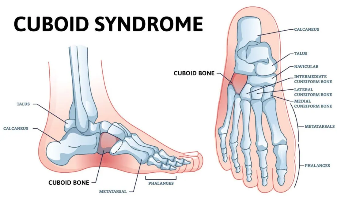
What is Cuboid Pain? Cuboid pain is not one of the most common foot complaints, but that’s why this blog is important for you to read. It’s a diagnosis that can be overlooked by healthcare professionals. The cuboid bone is a cube-shaped bone on the outside, lateral aspect of our foot – the pinky toe side of the foot. It lies halfway between our toes and heel. If you were to look at your foot from the side when you’re not bearing weight, it would be the bone that is at the highest part of your arch.

This area is called the lateral arch. The lateral arch is formed by the 4th and 5th metatarsal bones, which are the long bones that extend out to the ball of the foot that your bear weight is on. These two bones are connected to the cuboid bone by ligaments, and the bone forms the joint on the other side of the cuboid bone. The medial arch is the inner portion of the arch and is formed by the three metatarsal bones that connect to the midfoot. The medial arch is thought to be stronger than the lateral arch and can take more stress placed through this portion of the foot. This basic understanding is important to know as it plays a role in why you get cuboid pain. We will discuss the symptoms, diagnosis and cuboid syndrome treatment options in this blog.

What Happens When you Have Cuboid Syndrome Pain?

It is thought that the cuboid bone gets out of proper alignment with the calcaneus (heel) bone. This misalignment of the cuboid bone will cause pain in and around the joint. It can also be described as a midfoot sprain. It may also be referred to as cuboid subluxation.

Risk Factors for Cuboid Syndrome Include:

  • Trauma– ankle sprains, often described as an inversion ankle sprain. When this occurs, the ankle twists in such a way that the body weight rolls towards the outer side of the foot on the small toes’ joint side. The added weight in this area can cause displacement of the calcaneocuboid joint. Other types of foot injuries may also be a cause.
  • Foot biomechanics – individuals who overpronate or flatten their feet too much may be more prone to cuboid syndrome. It is thought that overpronation is the cause of cuboid pain for 80 percent of the population.
  • Repetitive stress – overdoing it, especially with weight-bearing activities, can also lead to this problem. Ballet dancers are especially prone to this injury because of the positions the activity requires their feet to be placed in.
  • Compensatory pain – one of the more common causes can be from pain elsewhere in the foot or ankle. We are very good at modifying our gait to help prevent pain. After a while, we may not even be aware we are doing it. If another foot problem requires us to walk on the outside of the foot to make it feel better, this may also be a cause. One example of this is plantar fasciitis. This will cause pain in the bottom of the heel, and it is common to compensate and avoid this pain by rolling your foot to the outside. If this is the case, it’s very important to resolve this pain first, and in many cases, this can fix the cuboid pain once the compensation stops.

What will my Symptoms be?

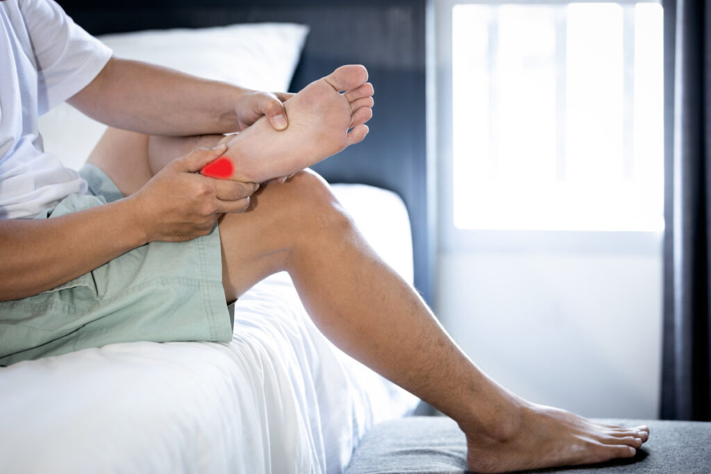
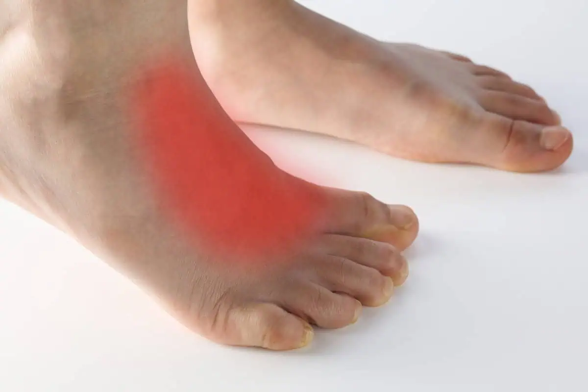
There will be pain on the outer side of the foot midway between the heel and the ball of the foot. The pain will occur when putting weight on the foot. It can be very severe and is usually a dull, deep pain. You may find you feel better when walking in shoes than barefoot as the support of the shoes may help with the discomfort.

How will the Doctor Diagnose Cuboid Syndrome?

The doctor can learn a lot from your explanation of where the pain is. Also, examination of the foot will be important as the pain will tend to be directly over the calcaneocuboid joint. There may also be some swelling. X-rays will also be taken to rule out fractures of the cuboid or adjacent bones. Other problems that could mimic cuboid syndrome include a fracture of the cuboid bone, tendonitis of tendons in the area called peroneal tendons. MRI or diagnostic ultrasound may be used to evaluate these other problems to determine what cuboid syndrome treatment will be the most effective.

Mysterious Pain on the Outside of the Foot? Here are the Treatment Options for Cuboid Syndrome!

  • Physical therapy – A physical therapist may be able to reduce pain in the cuboid area. This can also help to prevent the recurrence of cuboid syndrome.

This can be accomplished with

    • Balance exercises – By improving balance, you will be less likely to injure your foot as ankle sprains can be a common cause of cuboid syndrome.
    • Mobility exercises – To help regain full range of motion and get rid of any stiffness that may have occurred with cuboid syndrome.
    • Strengthening exercises – this will provide more strength to support the foot and ankle. Stronger muscles and tendons will provide more support to the foot and ankle and reduce the chance of cuboid syndrome.
    • Support of the cuboid bone – This may be accomplished by using padding underneath the cuboid bone area, over-the-counter arch supports may also help.
  • Orthotics – Custom-made inserts to go in your shoes may be the most important part of the treatment that your podiatrist can provide. This can give very precise support to that portion of your foot and lead to full recovery. It must be emphasized that orthotics may be used to treat the primary problem that created the cuboid syndrome. A very common example would be if you’ve been favoring your heel (plantar fasciitis) and this compensation has put added stress on the cuboid area. Walking differently and favoring your heel makes it feel better, but now you have cuboid pain. So by eliminating the heel pain with orthotics, the cuboid pain goes away.
  • Taping the foot and ankle – Taping the foot may also help support the calcaneocuboid joint area. This is especially effective in the early stage of the problem.
  • RICE – Rest, ice, compression, and elevation may also be used. These treatments will reduce inflammation in the area and promote healing. For some, resting the foot for up to 6-8 weeks may be necessary before full recovery and returning to regular activities.
  • Manipulation – This should only be attempted by a healthcare professional familiar with this technique. It is sometimes referred to as the cuboid whip. The technique involves placing the patient on their stomach with the knee on the involved side flexed. Then, with both hands gripping the foot, it is brought upwards and then thrust rapidly downward. This will allow the cuboid to pop back into its proper position.
  • Modifying activities– Cross-training may also be helpful. If you’ve been doing weight-bearing activities that involve a lot of side-to-side motion which may stress the cuboid joint area more, you may elect to take a break and bike,

As with many foot and ankle injuries, cuboid syndrome should be properly diagnosed and treated as soon as possible. This will help to speed up getting back to full activities. So, if you’re having a mysterious pain on the outside of your foot please consider the possibility of cuboid syndrome. We have skilled podiatrist who guide each of our patients to ensure a proper cuboid syndrome treatment is chosen that best suits the individual.

Call us today at our Fort Collins location (970) 329-8158, Broomfield location (303) 997-2795, Surgery Center (970) 329-8158, or use our online scheduling system to book your appointment.

The Perfect Read If You’re Considering Bunion Surgery Lapiplasty!

bunion surgery lapiplasty

Bunions are a deformity of the big toe joint that can make it difficult and painful to wear shoes. This affliction may be addressed by doing a bunion surgery lapiplasty correction when conservative options fail. If you’re noticing a bump on the inside of your big toe joint that is bothering you when you wear fashionable shoes – this blog is for you.

What is a Bunion?

A bunion deformity is caused by a misalignment of the bones in the foot. The first toe will angulate towards the second toe and the first metatarsal will angulate away from the second metatarsal.

Normally, the first and second metatarsal bones should be close to parallel to each other. However, with a bunion deformity, the angle between the two is increased. This will give the appearance of a large bump on the inside of your big toe joint. The actual bump can be from extra bone on the inside of what is referred to as the metatarsal head but more accurately the primary cause of the prominence is the angle between the metatarsal bones.

What is the goal of surgery?

The goal of surgery is to correct the angle between the first and second metatarsal bones. This correction brings the angle back to a normal anatomical alignment. We do this in such a way that there is no recurrence.

The History of Bunion Correction Procedures

Decades ago, many patients who had bunion surgery had the bump removed. They would use the soft tissue around the great toe joint to realign the toe. These procedures made for an easy recovery but resulted in minimal reduction of deformity and a very high recurrence rate.

bunions-infographic

Metatarsal Osteotomies

Next, foot and ankle surgeons started to do procedures with osteotomies on the first metatarsal bone to correct the deformity and remove the bump. An osteotomy means that the bone is cut and realigned or shifted to correct the angulation. There were two types of osteotomies; those performed at the metatarsal head (Austin or chevron) in order for the metatarsal head to be shifted. Otherwise, the osteotomies are performed at the base of the metatarsal bone. Those done at the base were for more severe bunions. These types of procedures involved significantly more lay up time. Whether the correction of the deformity is performed at the metatarsal head or the metatarsal base, they are considered traditional bunion surgery.

Next, surgeons determined that maybe the best surgical option was to fuse the first metatarsal cuneiform joint because this is considered an unstable joint. This is called the Lapidus procedure. It corrects the misalignment with a joint fusion. This can work very well but it includes a long lay time. Patients are not allowed to put weight on their foot for at least six weeks non weight bearing. There was also a risk of nonunion or delayed union of bone at the fusion site.

Bunion Surgery Lapiplasty

The bunion surgery lapiplasty procedure is a refinement of the Lapidus procedure. It is promoted as correcting the deformity in three different planes. The procedure also secures the unstable foundation which is believed to be the first metatarsal cuneiform joint. It also allows for early weight bearing. However, it is recommended that you be in a Cam walker so the bones are fully healed for at least six weeks. It is also promoted as having minimal recurrence rate. There are also claims that doing osteotomies of the metatarsal head have very high recurrence rates. Lapiplasty is promoted as having advanced fixation technology to fuse the joint. This technique uses titanium plating technology and these anatomically shaped titanium plates remain in the foot after surgery. Lastly, specialized instrumentation to make the results more precise and predictable are used.

Dr. Anderson’s Opinion of Bunion Surgery Lapiplasty

First, understand that I’ve been curious about this procedure but yet have not performed this procedure.

Here are some notes:

Recurrence Rates: In the marketing material for the Lapiplasty, they suggest a very high recurrence rate for patients that have an osteotomy of the metatarsal head. As a surgeon that has been doing bunion surgeries for 4 decades, I differ with this. Very seldom do we see recurrences. If surgeons were more like me, we would not have continued with these techniques.

They suggest that the correction is more precise because the bunion is being corrected in three planes and not two. In regards to this, yes, I can definitely agree with this; however, one would then assume that if the correction is not done in this manner, less correction was made, and recurrence rates were high – and on this I disagree.

With the Lapiplasty procedure, it is suggested that you will have less layup time. You’ll be able to bear weight within days and will ambulate in a Cam walker for 6 weeks. I’m not sure where the narrative comes from but most patients’ assumption of how long they are laid up is significantly more than the reality. The current way that we do most of our bunion corrections is a few days of non-weight bearing, then walking in a surgical boot or surgery shoe for up to 3-4 weeks. Patients can return to regular shoes in 3-4 weeks. Therefore the conventional approach to bunion surgery using a metatarsal head osteotomy is significantly less than the bunion surgery lapiplasty.

X-Rays of bunion surgery lapiplasty

X-Rays of Anderson Podiatry Center Technique

  • Majority of patients opt for our procedure due to successful outcomes
  • No pins left in the foot
  • Minimal chance of recurrence
  • You are back to wearing shoes faster

Hardware Complications: Currently we use small pins to fixate your bone with our bunion corrections. The advantage is that they are removed just weeks after, through a very small incision. Therefore, you don’t need to worry about potential pain from bone screws or plates that remain in your foot.

Marketing for Doctors: in the competitive medical world doctors are sometimes looking for an approach that sets them apart or allows them to market themselves. I have no problem with this but if you’re considering this procedure, please understand that there is motivation to do this procedure because the company that makes the instrument is marketing the doctors. You do the procedure and we will market your practice for you to generate more patients. Please consider this.

My Conclusion

I think the Lapiplasty bunion correction is a welcome addition for how foot and ankle surgeons correct a bunion. I don’t think it should be a one size fits all to be done on all bunions. I also think that it’s misleading to think that this procedure will lay you up less, as with many current approaches you may be back in shoes in 3-4 weeks, not 6 weeks. Many patients don’t know enough about bunion surgery to understand that indeed their bunion deformity may not require the procedures that require longer lay ups. In fact, the majority do not. I can agree that the technique used may lead to more consistent results, that’s assuming the conventional approaches have a lot of variation in outcomes. To summarize, you need to question the recurrence rates that they claim with conventional surgeries, and if it is worth the longer lay up times. If you are considering bunion correction please don’t hesitate to contact us for an initial bunion foot evaluation.

We have local foot clinics in the Fort Collins and Broomfield area with certified podiatrist who are experienced in bunion corrections. Watch out patient testimonial below to hear about the success, Phyllis is just one of many we have treated over the years.

For a full list of videos view our Bunion You Tube playlist.

Watch Videos

Plantar Nerve Entrapment: Causes, Symptoms and Effective Treatment

Plantar Nerve Entrapment

Heel pain is one of the most common foot complaints a foot specialist treats. For many, the term plantar fasciitis comes to mind when their heels start to hurt. Yes, plantar fasciitis is very common but other problems can be the cause of your heel pain. If your heel pain persists after months or years of self-treatment or treatment by your doctor, this blog is for you. You may be misdiagnosed and going down a dead-end pathway, you should consider if it is plantar nerve entrapment among other possible factors.

The Basics

The term plantar means bottom of the foot and the fascia is that strong connective tissue that runs from the ball of the foot to the bottom of the heel where it attaches. As your arch flattens, the fascia is pulled and pain can begin at the insertion on the bottom of your heel. However, it’s important to know that there are nerve branches in this same area that may be contributing to your pain.

The Tarsal Tunnel – The tarsal tunnel, much like the carpal tunnel in the hand, is an area where the nerve branches that have traveled down your leg go through to supply the bottom of your foot. The main nerve (See Diagram) is called the posterior tibial nerve.

plantar-nerve entrapment inforgraphic of nerve layout

As the posterior tibial nerve passes through the tarsal tunnel (which is composed of soft tissue structure) there can be excessive pressure on this nerve branch from those structures. We call this compression or a plantar nerve entrapment site. Following the posterior tibial nerve, it divides into three branches; one goes to the bottom of the heel and is called the medial calcaneal nerve. The other two are called the medial and lateral plantar nerve and supply the bottom of the foot. The medial branch supplies the bottom of the first and second, and a part of the third toe. The lateral plantar nerve supplies the fourth, fifth, and part of the third toe. Both of these nerves travel beneath a muscle called the abductor muscle. The tight fascial sheath beneath this muscle can compress (entrap) each of these two branches. If we follow the lateral plantar right after it goes beneath the abductor muscle, it sends a branch towards the bottom of the heel. This very small branch can be entrapped by the fascia of the quadratus plantae muscle. The branch is called the first branch of the lateral plantar nerve or Baxter’s nerve.

How do you know if you have entrapment of this plantar nerve which is a small branch of the lateral plantar nerve?

There will be subtle differences. This branch may cause more burning pain, tingling or numbness. The pain may be more dispersed in a larger area and not just where the plantar fascia attaches on the bottom of the heel. You may notice periodic tingling or numbness radiating towards your fourth or fifth toes. If you tap on the nerves in the tarsal tunnel area you may get a sensation like a funny bone sensation going down to your toes. This is called a tinel’s sign, and suggests the nerve in this area may be damaged from compression on the nerve.

What will your podiatrist do to test for plantar nerve entrapment?

Diagnostic Ultrasound – with every patient we see at Anderson Podiatry Center we evaluate your heel with ultrasound. This allows us to evaluate the soft tissue structures in this area including the nerve branches and the plantar fascia. If the fascia appears normal from this exam this will lead us to suspect that your heel pain may be more related to the nerves. It’s very important to know that there is no rule that says you only get one diagnosis. So, if we are treating plantar fasciitis because your ultrasound confirms it is damaged but you’re not progressing with our plantar fasciitis treatment, then this will raise our suspicions of another problem, potentially a plantar nerve.

What are the treatment options?

For The Patient – icing the area and taking autoinflammatory may help in the early stages and if the pain is mild. Make sure you’re wearing supportive shoes and you may consider trying over the counter arch supports. You may consider taking anti-inflammatory medication.

What will your foot specialist do?

Treatments may include using custom made arch supports, and orthotics. Additionally:

  • Laser Treatments – Our MLS laser system has been shown to be highly effective for treatment of nerve conditions.
  • Physical Therapy – ultrasound may have some benefit.
  • Cortisone Injections – A series of cortisone injections, 2 or possible 3 injections may be helpful. This is done with ultrasound guidance so the nerve can be located, and the injection delivered in the exact location.
  • Surgery – If the conservative options fail then surgery to release the pressure on the nerve from the soft tissue structures may be considered.

So, if you’re having heel pain, you may need to consider that it’s plantar nerve entrapment. Finally, you need to know that there are many well-meaning healthcare people whether it’s a physical therapist, family doctor, or people in the shoe industry that commonly assume if the bottom of your heel hurts then it’s plantar fasciitis. Understand that what you hear may be wrong and that is why the problem is persisting. Please consider revisiting your heel pain issue if you’re getting nowhere in your present course of treatment.

This Nerve Treatment Finally Fixed Her Heel Pain

Call us today at our Fort Collins location (970) 329-8158, Broomfield location (303) 997-2795, Surgery Center (970) 329-8158, or use our online scheduling system to book your appointment.

What Could It Be That’s Causing Your Ankle Pain Without Injury?

ankle pain without injury

The ankle joint is a very important joint that allows you to have proper mobility. When it starts hurting for no apparent reason and it’s slowing you down, you’re very likely feeling frustrated. You’ll start to wonder what I did and what can be done about it. In this blog I’ll give you some incites as to what the pain could be coming from. In many situations its it could be related to overuse but in other situation it may not be. This is why we think there should be a discussion about “ankle pain without injury?”, because this may stop a person from getting back to regular activities due to lingering joint issues.

The ankle joint– The ankle joint is one of the most important joints in your feet and ankles for normal ambulation. It’s the primary joint in the foot that allows the foot to move up and down which is very important for walking, running, and a multitude of activities. It has various ligaments and tendons around it that help to stabilize the joint and support your weight on the joint. The primary foot bone that sits inside the ankle between the tibia and fibula is called the talus. It is thought to be the hardest bone in your body. Why? Because all your body weight has to be placed on this small surface area of bone. Imagine how much wider your knee joint is compared to your ankle joint and you’ll begin to appreciate the strength of this bone inside the ankle joint.

Common causes of ankle pain

Problems associated with overuse or trauma

Osteoarthritis – another name for this is degenerative joint disease. This can occur for two reasons

  • Overuse – from years of activities that can cause damage to the joint, the wear and tear that will come over time
  • Trauma- old ankle injuries which may include a history of multiple ankle sprains. Every time you sprain your ankle there is the potential for damage to the cartilage inside the ankle joint. This repeated damage over time can create osteoarthritis state. This can then create joint pain.
  • Ankle fractures – If you have a history of an ankle fracture this can increase the likelihood of arthritis in the joint. In some cases, surgery may have been performed to reposition the bones in proper alignment and because of this the joint alignment may not be as precise as it was before surgery, and this may lead to more wear and tear to the joint cartilage over time.
  • Flatfeet – The type of foot you’re born with may also lead to ankle pain. If you have a foot that overpronates, flattens, this may also cause pain in the ankle joint. A flatfoot can lead to more wear and tear on the ankle. It may also lead to other problems including Achilles tendon pain which would cause pain behind the ankle. Achilles tendonitis would make the tendon painful to touch and may cause swelling. Another tendon that helps support the foot and ankle is called the posterior tibial tendon. It is located around the inside of the foot and ankle and may also get overused because of the flatfoot. This can cause pain and swelling on the inside of the ankle joint.

Problems not caused by overuse and trauma

  • Gout – Sudden ankle pain without injury can be from gout. The pain from gout can be very severe and can come on very quickly for no apparent reason. The ankle will become swollen, and heat and redness will be associated with it. The affected ankle will feel warmer to touch than the opposite ankle and will look more red. Gout can often be mistaken for an infection. Gout occurs as a byproduct of protein metabolism. Uric acid is a byproduct of that metabolic process. When Uric acid gets into a joint it’s recognized as being foreign to the body’s immune system. So, your body’s immune system begins to attack these uric acid crystals in the joint as if the crystals were bacteria. A medication called Indocin is frequently used to treat acute attacks. If the gout attacks keep returning further workup may be necessary to see if there is a related medical cause and potentially the use of medication to lower Uric acid levels in the body may be recommended.
  • Autoimmune disease – These may include rheumatoid arthritis and Lupus. An autoimmune disease will create inflammation to the ligaments and joint capsule around the ankle joint. This may also cause damage to these soft tissue structures leading to less support for the join and ultimately damage to the joint cartilage. With an autoimmune disease your body’s immune system will attack and damge these soft tissue structures. Medications are frequently needed to treat these conditions.
  • Ganglions – This is soft tissue fluid filled sacs that may originate from the ankle joint or from tendons sheaths around the ankle. If painful these are treated with aspiration of the fluid from the sac or surgical removal.

What can be done about ankle pain without injury?

If you find yourself experiencing ankle pain, consider some of the following suggestions.

  • If it’s very sudden and your ankle feels hot and looks red get immediate medical attention as most likely this may be a gout attack.
  • If there is no redness of heat consider RICE, rest, Ice, compression and elevation. You may also consider using non-steroidal anti-inflammatories NSAID, which may help.
  • If you have flat feet you may want to consider more support shoes or consider over the counter arch supports.
  • If the pain persists with your attempts, then consider seeing your podiatrist

What will a podiatrist do?

It starts with a good medical history from you as this can give us good insight into what it may be. X-rays and diagnostic ultrasound to look at the joint and the soft tissues around the joint may be needed. Examination of the foot and ankle by itself can tell us a lot simply by the appearance and location of the ankle pain without injury.

Options we consider

  • Orthotics– custom made arch supports may be recommended especially if the problem is related to flatfeet. Also, this treatment can be helpful if the joint is getting osteoarthritis from previous trauma, overuse or autoimmune diseases.
  • Regenerative medicine treatments- this involves using what is now referred to as human cellular tissue products. Currently we use umbilical cord or placenta cell products. These are injected into the joint in a clinic setting. Minimal down time is needed and there is minimal discomfort. This is usually a one-time treatment and we have had great success in helping with pain and prevent ankle surgery.
  • Laser treatment- MLS laser treatment can also help reduce the pain and inflammation in joint and surrounding soft tissues.
  • Surgery – This usually involves ankle scope surgery. Two small incisions are made to access the joint and through these incisions the joint and its structures can be visualized and damaged cartilage or soft tissue removed. Usually, you can get your foot wet within a few days and can bear weight within a few days. You can return to full activity relatively quickly with this technique. It’s also useful to have this done early if ankle arthritis is severe enough that ankle replacement surgery could be a consideration in the future.

I hope this blog eliminated some of the confusion that can happen when your ankle hurts without an injury.

Regenerative Patient Testimonial Video

Ankle Scope Patient Testimonial Video

Have arthritis pain on the top of your foot? How we avoid joint fusion surgery.

arthritis on top of foot

Feet are very important for daily function. Whether you are an active runner or like to go on walks for exercise, arthritis pain on the top of your foot can be an issue. The foot is very complex and we know that approximately one-quarter of all the bones in your body are in your feet. Halfway between the ball of the foot by the big toe and where you bear weight and the back of your foot whereby the ankle joint is the midfoot. There are multiple bones in this area that are cube shaped and comprise the arch. It’s a common area to get arthritis in the foot and ankle. These are the affected joints that may cause pain on the top of your foot commonly referred to as midfoot arthritis.

What are the common symptoms?

  1. A boney prominence in this area is associated with the arthritic joint.
    The prominence may rub against your shoe and cause pain.
    There may also be numbness, burning or tingling in the same area because the bone spurs are rubbing against nerves beneath the skin.
  2. Dull pain in the morning when you first bear weight.
    There may be pain and swelling on the top of the foot. After a few minutes of walking the pain may subside.
    Over time this pain may be more severe and prolonged hurting with every step you take

What are the causes of pain on the top of the foot?

The number one problem may be foot mechanics causing more wear and tear on these joints. This type of arthritis is called osteoarthritis. Some feet may be more prone to this issue. Therefore, you are born with the foot biomechanics that might make you more likely to get the problem. Oftentimes a foot type may put more forces through the inner side of the foot by the first and second toe joints. This will cause more stress at the two or three joints in the midfoot associated with these joints in the ball of your foot.

  • Trauma- there may be a history of trauma to the area. There may have been an old fracture of partial dislocation or sprain in the foot that now years later will create arthritis and pain.
  • Rheumatoid arthritis- pain from arthritis caused by autoimmune disorders such as rheumatoid arthritis.

What you can do for arthritis on top of foot?

  • You may need to get a larger shoe or one that is made up of softer material, so it does not rub against the boney spurs.
  • Get a stiffer soled shoe- if you’re getting dull pain in the area when you first bear weight in the morning this is a sign of joint arthritis so more support is critical.
  • Over the counter arch supports- using an over-the-counter arch support may also be helpful. This can add support to the shoes you have. In the elderly using devices such as a cane or walker may become necessary.
  • Choose different activities- If you run consider switching to hiking as this may cause less stress on the joints. You may also consider biking or swimming.
  • Lose weight- If you are overweight losing weight will put less stress through the joints and may reduce your pain.
  • Medications- consider using nonsteroidal anti-inflammatory drugs NSAIDS. These anti-inflammatory drugs may help with the pain but don’t correct the problem and long-term use may not be good for the joints.
  • Supplements and intermittent fasting – this may help reduce inflammation and pain.

What your podiatrist can do?

  • Orthotics – this is a common type of treatment recommended especially if you’re getting pain from the joints in your midfoot as this is an indicator of arthritis and more support and dramatically reduce your pain. This process involves getting a non-weight bearing digital scan of your foot so that an orthotic can be made to more accurately support your foot.
  • Cortisone injections – These may be helpful if there is nerve damage from the boney spur irritating nerves on the top of the foot.
  • Laser treatments – This may also be an option if nerve pain is an issue. Our MLS therapy laser is quick to provide relief with no downtime in comparison to surgery.
  • Surgery – Surgery to remove the boney spur may be recommended in some situations where there is minimal joint pain but simply pain from the boney prominence.

Treatments for Joint Pain from Foot Arthritis

As the problem progresses what was once simply a painful bump on the top of the foot may now become pain from the joint that the spur formation is associated with. Then joint pain can become very severe in this area. Fusion of the joint has always been the go-to surgical option. This may involve removing the joint or joints and then using plates staples or bone screws to fuse the joint. It can be successful but there are risks and the lay-up period is weeks of non-weight bearing.

Arthritis Pain on the Top of the Foot: Avoiding Joint Fusion Surgery

Arthritis in the midfoot, also known as midfoot arthritis, is a common condition that can cause significant pain and limit mobility. Many individuals with this condition experience discomfort on the top of the foot, often exacerbated by walking, standing, or wearing certain types of footwear. While joint fusion surgery is a traditional option for treating severe cases, several non-surgical treatments are now available that may help alleviate pain and restore function without the need for invasive procedures.

Surgical Options

When non-surgical treatments fail to provide sufficient relief, surgery may be necessary to restore function and alleviate chronic pain. Traditional joint fusion surgery, also known as arthrodesis, is a common surgical approach, but other techniques may be considered depending on the severity of the arthritis.

Joint Fusion (Arthrodesis)

Arthrodesis involves permanently fusing the affected bones together to eliminate movement in the arthritic joint, reducing pain. While this procedure is effective, it does limit flexibility in the foot and may increase stress on surrounding joints over time. Recovery typically includes a period of immobilization followed by physical therapy.

Joint Resurfacing or Cartilage Repair

For patients with less severe arthritis, procedures that focus on preserving joint function may be an option:

  • Cartilage Transplantation: If the damage is localized, healthy cartilage from another part of the body can be transplanted into the affected area.
  • Osteotomy: A surgical realignment of bones to redistribute pressure and alleviate pain without completely fusing the joint.

These alternative surgical procedures can help maintain more natural movement in the foot while still addressing the pain and instability caused by arthritis.

Non-Surgical Treatment Options

For those seeking to manage arthritis pain in the top of the foot without undergoing surgery, various non-invasive and minimally invasive treatments can offer relief. These include:

Regenerative Medicine Treatments

Regenerative medicine is a groundbreaking approach that focuses on harnessing the body’s natural healing processes to repair and regenerate damaged tissues. The following regenerative treatments have shown promise in treating midfoot arthritis:

  • Platelet-Rich Plasma (PRP) Therapy: This treatment involves extracting a patient’s blood, concentrating the platelets, and injecting them into the affected area. PRP contains growth factors that stimulate healing and reduce inflammation.
  • Stem Cell Therapy: Stem cells, typically derived from a patient’s bone marrow or adipose tissue, are injected into the arthritic joint to promote tissue repair and reduce pain.
  • Prolotherapy: A series of dextrose-based injections that stimulate the body’s natural healing response to strengthen and repair the weakened ligaments and joints.

These treatments can help improve joint function, reduce pain, and potentially slow down the progression of arthritis.

Joint Denervation

Joint denervation is an innovative technique that targets the nerves responsible for transmitting pain signals from the arthritic joint. By selectively disrupting these nerve signals, patients can experience significant pain relief without altering the structure of the foot.

  • Radiofrequency Ablation (RFA): This minimally invasive procedure uses heat generated by radio waves to disrupt nerve function, providing long-lasting pain relief.
  • Nerve Blocks: Injections of anesthetic agents can temporarily block pain signals and help determine whether denervation might be an effective long-term solution.

This approach is particularly beneficial for individuals who are not candidates for surgery or who prefer a treatment that does not involve altering the joint itself.

Choosing the Right Treatment for Arthritis on Top of Foot

Determining the best treatment option depends on the severity of the arthritis, the patient’s activity level, and overall health goals. While surgical interventions remain a viable solution for severe cases, non-surgical treatments like regenerative medicine and joint denervation offer promising alternatives for pain relief and improved mobility.

We do it differently at Anderson Podiatry Center!

  • Regenerative medicine treatments – for over 15 years we have been one of the leaders in podiatry using restorative medicine. We currently use placenta or umbilical cord products. These are injected into the involved joints. This allows the opportunity for your body’s natural healing processes to reduce inflammation and trigger increased growth factors to repair resulting in pain relief. We have had great success with this option, and it works well as these are not load bearing joints like a knee. With less load, the repair is thought to be improved. These are done in the office and patients return to normal activities in days.
  • Joint denervation – This is a process that involves blocking the pain signals to the brain by removal of a sensory nerve branch. There are four nerves in the lower leg that do not affect muscle strength. Any one of them can be safety removed and will not result in weakness. If a branch is removed, it is buried into muscle and patients are ambulating within days. The success rate has been very good and allows patients to delay and in most cases eliminate the need for joint fusion surgery.

As you can see we have multiple options to get you back to full activities and also feel we are well-versed to reduce the need for joint fusion surgery. View our patient testimonials below and when you are ready to meet with a local foot doctor in Fort Collins, give us a call.

Our foot clinic in Fort Collins or our other podiatric foot care office in Broomfield, Colorado  is staffed with compassionate, caring, and experienced staff. We will be glad to provide you with answers for arthritis on top of your foot.

Frequently Asked Questions (FAQ) for Arthritis on Top of Foot

    • What are the early signs of midfoot arthritis?
      Common early signs include pain on the top of the foot, swelling, stiffness, and difficulty walking or wearing certain shoes.
    • How can regenerative medicine help with midfoot arthritis?
      Regenerative treatments like PRP therapy, stem cell therapy, and prolotherapy promote healing, reduce inflammation, and improve joint function without surgery.
    • Is joint denervation a permanent solution?
      Joint denervation can provide long-lasting pain relief, but results may vary. Some patients may require repeat treatments over time.
    • What are the risks of joint fusion surgery?
      Potential risks include loss of flexibility, increased stress on surrounding joints, prolonged recovery time, and possible complications such as infection or improper bone healing.
    • How do I know which treatment is right for me?
      A podiatric specialist can evaluate your condition and recommend the best treatment based on your symptoms, severity of arthritis, and lifestyle.
    • Can I still be active with midfoot arthritis?
      Yes, with proper management, including physical therapy, supportive footwear, and non-surgical treatments, many people with midfoot arthritis can maintain an active lifestyle.
    • How long does it take to recover from joint fusion surgery?
      Recovery can take several months, including immobilization and physical therapy. Full recovery may take up to a year, depending on individual healing rates.
    • Are there lifestyle changes that can help manage midfoot arthritis?
      Yes, maintaining a healthy weight, wearing supportive footwear, and performing low-impact exercises like swimming or cycling can help manage symptoms and reduce stress on the joints.

Discover the Difference Between Corns and Calluses

difference between callus and corn

Read This and You’ll Know the Difference Between Callus and Corn…and What to Do!

A Callus and a Corn are similar and are among the most common foot problems. However, in this blog, we will discuss the differences and what can be done to reduce or eliminate these painful conditions. The symptoms of corns and calluses can vary from a minor irritant to severe pain because of the difference between callus and corn location on the feet. Of all the foot ankle problems, corns and calluses can be the most irritating for some.

What are Calluses?

A callus is an area of skin build up that becomes hard and this dead skin becomes painful. The layers of skin become thickened. Calluses tend to be larger than corn and often don’t have a painful core in them which is more common with corn. Calluses also tend to be on weight bearing surfaces of the foot especially on the bottom. There are different types of calluses. Some are very diffuse and not very deep, therefore not too painful. The most painful are deeper and may have a central core of dead skin that is very sensitive to pressure. This type of callus is most painful. Both calluses and corns can be painful to touch.

What are the symptoms of calluses on feet?

  • Thickened, rough, and hardened patches of skin.
  • Usually appear on the soles of the feet, especially on the heels or balls of the feet.
  • Often larger and less defined than corns.
  • Can be less sensitive to touch compared to surrounding skin.
  • Generally not painful, but may cause discomfort if they become too thick.

Causes of Calluses Include:

  • Repeated friction and pressure from activities such as walking, running, or wearing tight or ill-fitting shoes.
  • High heels or shoes with thin soles that do not provide adequate cushioning.
  • Certain foot deformities, like bunions or hammertoes, which alter the way pressure is applied to the feet.
  • Wearing shoes without socks, leading to more direct friction.

What are Corns?

A corn tends to be smaller than a callus but is more likely to have a painful core in the center of it. A corn tends to be composed of a buildup of thickened skin much like callous. However, you can have another type of corn called a soft corn that may appear between your toes especially between the fourth and fifth toes in the 4th webspace. Generally, a corn is located on the toes.

What are the symptoms of corns on feet?

  • Small, well-defined, and often round thickened areas of skin.
  • Typically found on non-weight-bearing parts of the feet, such as the tops and sides of toes or between toes.
  • Can be hard (with a dense core) or soft (usually found between toes and kept moist by sweat).
  • May cause significant pain or discomfort, especially when pressed.

Causes of developing corns on your feet include:

  • Excessive pressure or friction from tight or poorly fitting shoes.
  • Toe deformities such as hammertoes or claw toes, which cause the toes to rub against the shoes.
  • Abnormal gait or walking patterns that increase pressure on certain areas of the feet.
  • Repeatedly wearing high heels, which shift weight to the front of the feet.

How Can You Prevent Suffering from Corns and Calluses?

A callus tends to be located in an area where there is excessive weight being placed, especially on the ball of the foot. We bear weight on the ball of the foot on the metatarsal heads. There are five metatarsal bones that take most of the weight on the ball of the foot. From improper biomechanics of foot structure, extra weight may be placed on one of these bones and the body’s reaction to that pressure is to build up the skin which we call a callus. A structure issue that some people are born with is called a Mortens Foot. With a Mortens foot, the first metatarsal which connects to the great toe may be shorter than it should be. When this occurs, your foot will put more weight than it should on the adjacent second metatarsal head causing a callus to form.

An example of a corn is when it’s associated with a hammertoe. This can occur on any of the toes but is rarely seen on the great toe. Oftentimes the small toe, 5th digit is hammered and curved towards the adjacent 4th toe. This is probably the most common type of hammertoe to develop a corn. A corn will then be created on the side of the toe that then rubs the shoe. In some cases, the rubbing of the 5th toe against the 4th toe may also create an irritation and therefore a corn will develop. The second digit is also a common place for corn to appear. It may be on the top of the knuckle of the toe and the tip if the digit.

Ideas For Treatment of Corns or Calluses at Home

Footwear:

  • Choose properly fitting shoes with adequate cushioning and support. Shoes should not be too tight or too loose.
  • Avoid high heels or shoes with thin soles that do not provide sufficient padding.
  • Consider using shoes with a wide toe box to reduce pressure on the toes.
  • Wear moisture-wicking socks that provide cushioning to reduce friction.
  • Avoid walking barefoot on hard surfaces.

Foot Hygiene:

  • Keep feet clean and moisturized to prevent dry, rough skin that can lead to calluses.
  • Regularly exfoliate feet to remove dead skin.

Protective Pads:

  • Use over-the-counter pads or cushions to protect areas prone to friction and pressure.
  • Custom orthotics may help distribute weight more evenly and reduce pressure on specific areas of the feet.

Activity Management:

  • Take breaks from activities that put excessive pressure on your feet.
  • Alternate between different types of footwear to avoid constant pressure on the same areas.

infographic of callus corn epidermal difference

What Your Podiatrist May Recommend Based On The Difference Between The Callus and Corn

For calluses on the ball of the foot, custom made inserts (orthotics ) may be recommended to reduce the buildup of skin and slow down its recurrence.
Periodic debridement may be recommended as this provides the option of having the podiatrist trim the tissue down. This may be advised every 2-4 months. This is also a common recommendation for those with diabetes. Tolcylen soaks can also be recommended. This is a new product that we currently sell though our office that is getting rave reviews. By using this soak on a regular basis, it gradually eats away at the dead tissues and also moisturizes your skin. It’s been exciting to introduce to our patients.

Below are some images that show the improvement our patients received when using Tolcylen.

tolcylen-therapeutic-foot-soak-15pouches-30days

tolcylen-therapeutic-foot-soak-5pouches-10days

tolcylen-therapeutic-foot-soak-5pouches-5days-sideview

tolcylen-therapeutic-foot-soak-5pouches-10days callus removal

Surgery is seldom, however it can be an option when conservative measures fail. Surgeries involve addressing the issues with the bones that we bear weight on to reduce the weight bearing on that specific bone.

Treatment of Corns and Callus

There are multiple options when it comes to the treatment of corns.

  • Pumice Stone: This can be used to periodically trim down the buildup of skin.
  • Inserts (Orthotics): To know if shoe inserts may help examine your feet. Shoe inserts may help if the callous in on the ball of the foot. The inserts may also reduce the weight load on the callus and help reduce the pain
  • Soaking Your Feet in Warm Water: This can be useful to soften the tissue especially before the use of a pumice stone.
  • Wearing Socks: It may be important to consider wearing socks frequently if you don’t as the padding from the soft sock may help.
  • Shoe Gear: To prevent corns and calluses selection of shoe gear may be important.

Now you know the difference between callus and corn, you should be able to determine what treatments to consider in your plan. When speaking to your local podiatrist in Fort Collins, Loveland, Broomfield or the Denver, Colorado area consider the options we have outlined above. Feel free to reach out to your qualified, experienced and certified podiatrist at our clinical location in Broomfield for advice or schedule an appointment.

How Laser Treatment Improves Your Ability to Get Rid of Foot and Ankle Pain!

laser treatment for foot pain MLS

At Anderson Podiatry Center, we like to bring you cutting edge technology. We feel it will allow us to deliver the best results for your foot and ankle care – and laser treatment is one of these technologies. We use the MLS laser system which brings the latest advancements in laser technology for your treatments. Laser is a treatment that is offered if conventional treatments are not working. This blog will introduce you to the laser treatment for foot pain option for foot and ankle issues.

How Does Laser Treatment for Foot Pain Work?

Our MLS laser therapy uses deeply penetrating light to help repair your body’s tissues. It is a “multi wave lock system”. MLS, that uses specific wavelengths of light. One wavelength penetrates closer to the surface and is continuous. Its primary function is to reduce swelling and inflammation. The second wavelength is pulsed and reaches deeper into the tissues and nerves for pain relief. Compared to earlier generation lasers, these lasers penetrate well below the skin layer (2-3 cm) allowing for more successful results. The laser works by stimulating your body’s own reparative processes. It very specifically helps the mitochondria in each of your body’s cells to do a better job. The mitochondria is an area of each cell that creates energy for the cell, repairs the cell, and eliminates waste from the cell. In this process three things occur:

  • Reduced Inflammation: The laser treatment can reduce inflammation and help speed up the reparative processes. This anti-inflammatory effect results with pain relief. This process also reduces swelling.
  • Positive effect on Pain Receptors: It also helps to reduce pain because of its effect on pain receptors. While your body’s healing processes are accelerated, the laser also helps to reduce pain.
  • More Blood Flow: The laser increases the blood to the targeted area. This aids in the healing processes which accelerates healing.

What is Treatment Like?

During this process, the laser light will be applied to the area being treated. There are a few things you’ll notice throughout the process:

  • You may feel a warming sensation, but the treatment is painless and there are no side effects.
  • The treatments can last for 15 to 30 minutes depending on how many areas are treated.
  • Generally, 6-12 treatments are recommended and are done 2-3 times per week.
  • You may begin to feel improvement after the first one or two treatments.

What Conditions Can Lasers Be Used For?

There are various reasons why we use laser for foot pain. Most commonly, we use laser in conjunction with surgery to accelerate healing and reduce swelling. Lasers can reduce scar tissue after surgery on joints to reduce stiffness. It can also accelerate the repair of nerves when we do nerve decompression surgeries to reverse the symptoms of neuropathy and restless legs. It is also used in conjunction with other treatment modalities for common foot and ankle conditions such as:

  • Heel Pain (plantar fasciitis): This is one of the most common foot complaints.
  • Neuroma Pain: This pain is in the ball of the foot which causes compression of a nerve branch in the ball of the foot.
  • Achilles tendonitis and other types of tendonitis.
  • Joint Pain: Laser has also been used to reduce the symptoms of arthritis in foot and ankle joints.
  • Neuropathy: This is a condition that causes burning, tingling and numbness in the foot and ankle.
  • Foot and Ankle Ulcers: This can speed up the repair of ulcer common in the diabetic foot.
  • Restless Legs: We have also used laser in conjunction with ESTIM electrical stimulation to reverse the symptoms of restless legs.
  • Foot and Ankle Injuries: It can speed up recovery from soft tissue injuries such as foot and ankle sprains. It can speed up the repair of damaged tissue.

In general, laser technology has allowed us to offer more options and to optimize the results we can offer our patients. If you’re frustrated with your foot and ankle issues, it’s something you may want to consider laser as a treatment option.

Testimonial from our patient who received laser treatment for foot pain!

roy-achilles-tendon-pain-laser-treatment-pain-mls


Cutting Edge MLS Laser – Doctor/Patient Feedback

MLS doctor testimonials laser treatment ankle pain

Understanding How Laser Therapy For Plantar Fasciitis Works! Is It Right For You?

Laser Pain Therapy Plantar Fasciitis

Heel pain is usually caused by plantar fasciitis, and laser therapy is a common noninvasive treatment we recommend. Many suffer from plantar fasciitis because it’s a very common problem. If you have heel pain, there are many treatments you will hear about.

How Do You Know If You Have Plantar Fasciitis?

Plantar fasciitis usually causes pain in the bottom of the heel bone. Often when you first get up in the morning is when it’s most painful. It’s thought that as you step down, the fascia, which is a strong fibrous band of connective tissue that extends from the ball of the foot to where it attaches to the heel bone, will be pulled. This is thought to cause damage to the fascia as the pulling may create microscopic tears where it attaches to the heel bone.

What is Important For You to Understand Before We Discuss How Laser Therapy for Plantar Fasciitis Can Help ?

Additional treatment methods for plantar fasciitis pain are

  • Stretching – Stretching is commonly recommended and although it may help for many, it may give a band aid approach, often not permanently resolving the problem.
  • Orthotic Devices – Custom made inserts may be very useful to help support the foot and decrease the pulling of the fascia from the heel bone. This pulling is thought to be the root cause of the problem.
  • Cortisone Injections – Cortisone can give temporary relief and is seldom a permanent solution.
  • Restorative Medicine – This form of treatment is highly successful and involves using human cellular tissue products such as stem cells. These are commonly derived from the placenta or umbilical cord to be injected into the area to repair the damaged tissues.
  • Surgery – When conservative measures fail, surgery may be performed. The surgery is done endoscopically through small incisions. A portion of the plantar fascia is released allowing it to be lengthened. Patients ambulate immediately in a cam walker for approximately 3 weeks.

Laser Basics

Laser treatment has been an important part of therapy for plantar fasciitis at Anderson Podiatry center for many years. It is used to help treat many foot and ankle problems. The advantage is that laser therapy offers its safety with no known side effects and is minimally invasive. The treatments are not painful and the lasers we use are robotic cutting-edge lasers. Our lasers are class IV level laser therapy lasers. The laser beam is set to trace over the area needing treatment. Our lasers are FDA approved cold lasers meaning there will be no heat generated or low level intensity during the treatment. The other class of lasers are called hot lasers. These lasers are used to destroy tissue such as warts or cut through tissues in surgery.

To understand how our cutting-edge lasers work we must first understand the mitochondria. Our lasers are deeply penetrating allowing for much more therapeutic value then most lasers. If you are up to speed in health, you may have heard about the mitochondrial. When you investigate a cell in your body, whether it’s a bone cell, muscle cell, or in this situation, the cells in the fascia of the foot, there is an area inside the cell called the mitochondria. The mitochondria is very important as it creates energy for the cell, repairs the cell and gets rid of toxins in the cell. When asked how a laser can help, you may often be told it gets rid of inflammation in the area, it increases blood flow and helps repair the tissues. These may all be true, however research shows that it all gets back to improving the function of the mitochondria.

Currently you may have heard that other things can improve the function of your mitochondria. This may include fasting, exposure to sunlight and getting good sleep. So, think of laser as a therapy that can add to your mitochondria to repair your body. In this situation it’s your damaged fascia.

How Is Laser Therapy for Plantar Fasciitis Performed?

Generally, laser treatments involve 6 to 12 treatments. With each treatment you will gradually experience reduction of pain. How fast and how much relief you get can vary. Laser is frequently used in combination with other treatments such as orthotics, restorative medicine etc. And for many, it does NOT become a maintenance program where you periodically need to keep coming in for more treatments.

We have had great success with laser therapy for plantar fasciitis because it is a low invasive and pain free treatment. We like this approach as we feel it’s really getting to the root cause of the problem by promoting your body’s own processes to heal the fascia which has been damaged. Our multilevel approach of using laser in combination with other treatments such as orthotics and restorative medicine, allows us to have a high success rate because we were attacking the root cause.

by Dr James Anderson, DPM, ABPS, FACFS, FAENS

Call us today at our Fort Collins location (970) 329-8158, Broomfield location (303) 997-2795, Surgery Center (970) 329-8158, or use our online scheduling system to book your appointment.

What You Need to Know About Foot Surgery for Bunions and Three Ways They are Corrected!

foot-surgery-for-bunions

Bunions can be a painful foot condition for many. They can become painful and make finding a comfortable pair of shoes to wear very difficult. Foot surgery for bunions should be not only be considered for the asthetical value, but the overall bony health and balance of the body.

What is a Bunion & What Causes a Bunion?

There is a primary problem that creates a bunion, which starts as the big toe begins to anglalate towards the smaller toes. The metatarsal bone at the base of the digit moves away from the rest of the foot causing an increased angle. This increased angle is between the first and second metatarsal bones. The results are a hard bony prominence at the base of the big toe. The medical term for this condition is Hallux Valgus.

The primary cause is thought that poor foot mechanics may be the main cause. Too much weight may be placed beneath the great toe joint, and it changes the pull of the tendon around the great toe which starts the process. Also, wearing shoes such as high heels that are pointed or too tight may also be a cause.

Non Surgical Treatments

When it comes to bunions, there are multiple non-surgical options to tryout:

  1. Change in Shoe Gear – Try to avoid shoes that are too tight and purchase shoes with a larger toe box
  2. Padding – Placing more cushion on the area may also provide temporary relief and the use of anti-inflammatory drugs
  3. Orthotics – Custom made shoe inserts called orthotics may also be useful

Surgical Treatment Option for Bunions

For many, the surgical correction becomes the treatment of choice. When it’s difficult to wear the majority of shoes this may be the appropriate treatment.

We can discuss three basic approaches to correcting the bunion. To begin with the problem lies in the angulation of the first metatarsal away from the second metatarsal bone. This is what causes most of the bony prominences that rub against the shoes. This angle needs to be reduced so the two bones are more parallel. By accomplishing this angle correction, it is easier to straighten the big toe. Many years ago, bunion surgery had a bad reputation because doctors would simply remove the bone bump and not address the angulation of the bone. Thus, for many, this only offered part-time relief.

How is the angle corrected? It can be done in three general ways:

  • By shifting the bone close the great toe joint
  • By shifting the bone by the base of the metatarsal bone closer to the instep
  • By removing a portion of the great toe joint

It is also important to note that what procedure the foot and ankle surgeon choses is based upon the severity of the bunion deformities and the patient’s age.

Three Types of Foot Surgery for Bunions Performed

  1. There is the option of shifting the bone by the great toe joint. The vast majority of toe time, this procedure is performed at Anderson Podiatry Center. The technique is used for more mild or moderate bunions. The recovery from this is usually wearing a surgical shoe or Cam walker for 3-4 weeks and just a few days of non-weight being in the beginning.
  2. The option of shifting the bone at the base closer to the ankle usually requires more lay-up time of weeks after surgery, usually 4-6 of off- weight bearing. This procedure is reserved for more severe deformities and may only comprise ten percent of the bunion corrections that we do.
  3. This final option is to remove a portion of the great toe joint. This can work very well in the senior population over 65 years of age. There is very little risk and has been performed successfully in patients in their 80’s. For all these surgical options, the affected foot will need to be elevated after surgery for several days and weight bearing can resume within days or weeks depending on what surgical approach is performed. Physical therapy may also be recommended during the surgical recovery. When you can return to normal activity level will also vary.

Below, I have a few notes in regards to bunion surgery and occurrence.

Surgical Setting: These surgeries are performed on an outpatient basis using local anesthesia and IV Sedation. Depending on the circumstance, both feet may be done at once.

Recurrence Rates: The possibility of recurrence of the bunion is very minimal especially if the foot and ankle surgeon is shifting the first metatarsal bone, and not simply removing the bunion bump.

Why Early Treatment May be Advantageous

Foot Surgery for bunions should be carefully considered and should not be done for cosmetic reasons. Once there is pain and shoes are becoming a challenge, surgery before the bunion becomes more severe may be best. Why? Simply because of lay-up time. This is something few patients are aware of. Many assume it may be weeks of not bearing weight on the foot. This is not the case for many patients. The majority of corrections we perform allow you to place weight on the foot within a few days.

Call us today at our Fort Collins location (970) 329-8158, Broomfield location (303) 997-2795, Surgery Center (970) 329-8158, or use our online scheduling system to book your appointment.

We can find a solution for you!

Bunion pain relief ! Karen is back to wearing the shoes she loves. She shares her experience.

Bunion Surgery Success! Tammy Shares Her Story

Susan’s shows off her shoes she can now wear . Bunions fixed !

Bunion surgery on both feet! Kari shares her honest story of what it was like for her.
Bunion Surgery – Kari

Do I have Fingernail Fungus? What’s The Best Form of Laser Treatment?

fingernail fungus laser treatment

Fingernail fungus can be a very troubling problem and getting laser treatment is what we recommend for this embarrassing condition. Although it’s most common on toenails, it may also be present on the fingernails. The embarrassment of finger fungus may be more common as it can be hidden when wearing your shoes. Fungus likes a warm moist place to grow and that is thought to be the reason for it to be more prevalent.

What does fingernail fungus look like?

  • The nail will become discolored such as a yellow discoloration, but the nail may also have white discoloration.
  • Thickening of the finger nail.
  • Deformation and curing in on the edges of the nail may cause the nail to become ingrown.

What Causes Nail Fungus Infections?

  • Fungus likes a moist place to grow and if your hands are in water a lot this may promote its presence.
  • Trauma to the nail may also start the process, and the trauma may cause a fissure in the nail and this allows fungus to enter the nail.
  • Your immune system – We need to understand that for a majority of people, it’s likely that the immune system is to blame. That is why one spouse may have it for years and the other never gets it.

What Can You Do to Avoid It?

  • It’s best to make sure you dry your hands well after bathing or washing them. Also, avoid wearing gloves for long periods of time as the gloves may trap moisture inside creating a better environment for fungus to grow.
  • Stay healthy – any medical condition that affects your immune system may make you more susceptible to nail fungus. A common example of this is diabetes.
  • Nail polish – Avoid nail polish as this is unhealthy for the nails and may lock moisture beneath it.
  • Finally, be kind to yourself. It’s likely not your fault. Many times, I tell many patients that they have less control than they think as the immune system they inherited just doesn’t fight fungus well. The fact is that many patients with this condition have a family history of fungus on their nails.

Fingernail Fungus Treatments can Include Three Options.

Topicals – Applying over the counter topicals may be your first step. If you catch it very early and it’s not too severe this may be your solution. However, out of all the options, it’s the least successful. So early detection and treatment is important for this option to work. It’s very difficult for topicals to penetrate a nail and that is why the success rate is low. I often am asked if removal of the nail along with treatment as it grows out will work. I’ve seldom seen this be effective. I feel this is not a good option as the removal itself may traumatize the nail and cause it to be deformed.

Oral medication – Lamisil (Terbinafine) is an oral medication. I consider this the second most successful treatment option. At Anderson Podiatry Center, we seldom prescribe antifungal oral medication because of the potential for liver toxicity and if we do, we get a liver panel order before and during treatment to ensure no liver damage is present before starting or during treatment. Many reports show a 40-60-percent success rate with the oral option.

Laser treatment – Laser is considered the most successful fingernail fungus laser treatment option. We have been performing laser treatments for 15 years and we’re the first to bring the treatment option to the Rocky Mountain region. We have a lot of experience! Laser works on the concept of heat. It penetrates heat deep within the nail and this is what destroys the fungus. Our pinpoint laser system pulses the heat very rapidly so most patients have no discomfort when treatment is done. There are also no known side effects and no symptoms after treatments are performed. As fingernails take four to six months to grow back. From our experience, we have found that a minimum of three treatments are recommended to get the maximum benefit of laser treatment for fingernail or toe nail fungus. The laser treatment cost is lower than expected and takes no more than 30 minutes per session. The cure rate for a fungal nail infection is the highest of the three options. We also offer antifungal nail polish in our offices to compliment your treatments.

fungal nail treatment using the laser pinponte system

Finally, you may have questioned why a podiatrist is doing a blog on fingernail fungus. The answer is we have had a lot of experience treating toenail fungus and feel we can offer the same for fingernails!

However, for us to proceed with our treatments, we must have an order and referral from your primary care doctor. So, if you’d like to better understand this process please email or call us for more information or request an appointment!