Image Menu canvas
Book Now

General Podiatry Archives - Page 6 of 17 - Anderson Podiatry Center Anderson Podiatry Center

General Podiatry Archives - Page 6 of 17 - Anderson Podiatry Center Anderson Podiatry Center

How Do You Know If You Have a Foot Ligament Injury?

foot ligament injury treatment in fort collins colorado

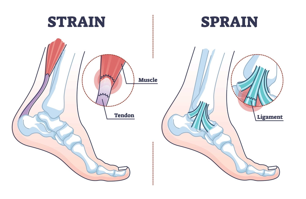
Foot ligament injury is a common problem in the foot and ankle. A ligament is a strong fibrous tissue whose function is to stabilize joints. A ligament is the tissue that connects the bones that form the joints. Ligaments can be injured from overuse but are more commonly injured from trauma. Ligament injuries are referred to as foot and ankle sprains. And out of all the possible ligament injuries, ankle sprains are the most common.

How Do I Know If I Have A Foot Ligament Injury In My Foot?

  • You may feel or hear something pop or snap
  • You may have pain and swelling in the area of injury
  • Don’t assume if you can walk on your foot or ankle that the ligament is not injured
  • It hurts when you put weight on your foot.

How Do We Know If The Ligament Is Injured?

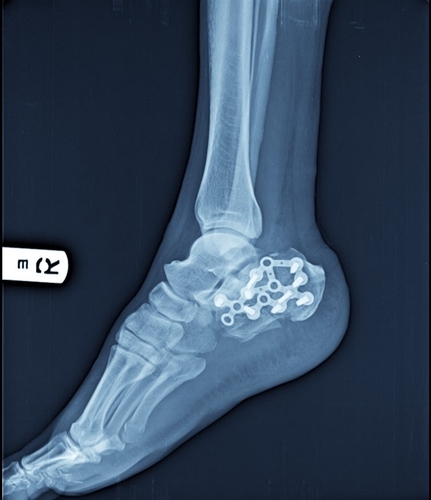
It starts by taking x-rays in most cases to rule out bone injuries. X-rays may also be used to see if a joint has more motion in it than normal. These are called stress views and your local podiatrist will advise you after reviewing the results. This can indicate that the ligament is no longer stabilizing the joint, and this is sometimes done to evaluate ankle sprains. An MRI may be ordered. This can help the doctor know if it is a ligament that is injured, and the severity. There are three different grades of tearing depending on the severity of the injury. Grade 1 is the mildest and means most of the ligament is not torn. Grade 2 indicates that 40-60 percent of the ligament is torn. A large tear is grade 3 and this indicates that the tear is completely through the ligament.

Treatment Of A Foot Ligament Injury

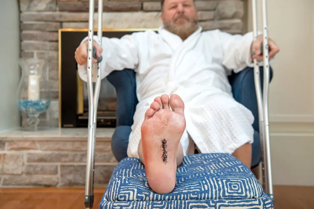
The options for treatment of a foot ligament injury vary depending on the severity. The most severe tears may need immobilization for weeks using a Cam walker. Some doctors may even suggest surgical repair to reconnect the torn ligament ends to allow them to better heal. With a more minor tear such as a grade 1, resting, icing, and elevation with gradual return to physical activities may be all that is needed.

Ankle Sprains

Of all the potential ligament injuries of the foot and ankle, injuries to the ligaments of the ankle are most common. It’s important to consider if it’s the first ankle sprain you’ve had, or if you have a history of repeated sprains. If it’s your first one and it’s associated with severe pain and swelling, a sense of something popping or giving out is important to note. In this situation, you should be evaluated by a foot and ankle specialist. With a severe sprain, a complete tear would mean that immobilization in a Cam Walker may be recommended. This will allow the ligament ends to be in closer approximation and provide a better chance of full repair. Without proper treatment, you may now be left with an ankle joint that is chronically unstable. Without proper treatment from the beginning, the instability of the ankle may become chronic. In some cases, this might require surgery to stabilize the ankle. Ankle sprains tend to be the most under evaluated and undertreated foot and ankle injury. Patients often go to an emergency room or urgent care facility. If x-rays rule out a fracture, little after care may be given.

What You Can Do To Prevent Ankle Sprains?

  • Consider wearing high top athletic shoes if you’re playing basketball
  • Use an over-the-counter ankle brace
  • Consider custom made orthotics as these give your feet better feedback regarding foot placement
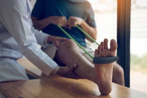
  • Do ankle exercises and consider trail hiking or running to build up muscle strength. This may help support your ankle joint more

So, it is important to understand that no matter where the foot ligament injury (ankle or foot) is, a ligament tear is to be suspected. Just because you can walk on it, and just because it’s not fractured doesn’t mean aggressive treatment wouldn’t be a good idea. As it’s often been said, a sprain can be almost as bad as a fracture. Finally, it is always better to error on the side of caution and consult with you podiatrist if you have concerns about a possible ligament injury.

Dr. Anderson, the founder of Anderson Podiatry Clinic, DPM, ABPS, FACFS, FAENS

Flat Feet Pronation: To Better Understand Overpronation and Why Flat Feet Are an Issue!

flat feet pronation

So, you’ve heard the words flat feet pronation, but what is it? Pronation is a commonly used word to describe motion in your foot that naturally occurs. It’s a term you may have heard from a shoe salesman or if you’ve read about a potential cause of injuries during your athletic endeavors. Knowing this, let me now contrast flatfeet and pronation.

Putting it All Together – Keep Reading to Better Understand How Well Our Feet Are Engineered!

By our definition, flatfeet refers to the appearance of a foot with minimal to no arch. The footprint of this foot would show very little arch. This type of foot could have a foot that is rigid, meaning even when not bearing weight the arch of the foot is absent. Very few have a flat foot that is this severe. Most are flexible, and when non weight bearing an arch is present. It is important to know that most flatfeet are thought to be acquired.

Another definition to keep in mind is pronation.This refers to the motion that occurs on your foot. We were all built to have this motion occur. The biomechanics of the foot is remarkable when it’s fully understood. The motion of pronation is complex as it means your foot is moving in all body planes. It would be like a universal joint on a car. As the entire foot rolls in, something remarkable occurs. The bones of the foot that support the arch become more flexible meaning they align is such a way to provide more motion which equals more shock absorption. This shock absorption helps when you walk or run to reduce the stress of impact on the feet, knees, and hips. Again, if your feet are the suspension of a car, pronation absorbs the shock of your body weight coming down on the foot. So some pronation is necessary, but too much is not a good thing as it causes stress on the foot, ankle, knees, and hips. It may also be a cause of back pain.

What Does Flat Feet Pronation Look Like?

Some feet will have a heel bone that rolls in too much. Normally, when you are standing, your heel should be close to perpendicular to the ground or slightly rotated inward (pronated). Too much rolling inward of the heel means you’re overpronated.

Other feet will overpronate with more of the motion occurring in the arch. In this foot type, the heel may remain close to perpendicular to the ground and the arch is flat. You may pronate more in the rearfoot or more in the arch, or a combination of both. So remember that pronation is not a bad thing as it is good to have some pronation but not too much.

What Problems Can be Related to Over Pronation?

  • Knee Pain – This can occur from overpronation causing too much inward deviation at the knee joint.
  • Back Pain – It is thought that overpronation will cause the pelvis to tilt forward causing symptoms in the lower back.
  • Bunions – This painful foot deformity is thought to be related to excessive weight beneath the great toe joint.
  • Plantar Fasciitis This is the primary cause of pain beneath the heel bone.

Treatment of Flat Feet Pronation

correction of flat feet pronation using orthotics

  • Orthotics – This has been the primary treatment for patients with flat feet pronation for most podiatrists. It’s often times not enough to shop for more supportive shoes. The process involves taking a non- weight bearing impression of your foot, placing it in a position it should be in, not the position it is in while standing. Moving the orthotic from shoe to shoe allows you to take the customized support you need with it. So, the impact of the shoes is neutralized.
  • Motion Control Shoes – When shopping for shoes you should ask for a motion control shoe. As an example, running shoes can be constructed with a lot of motion control built into the shoe or very little.The heel counter that cups the heel should be rigid. This is something to look for in a shoe if you overpronate.
  • Stretching – For some this may be helpful as a tight Achilles tendon may be associated with a flat foot.
  • Foot Exercises – There may be exercises that can be performed to increase the strength of muscles. This may have limited effect on more severe flatfeet.
  • Surgery – In certain situations surgeries to correct the flatfoot may be considered. The severity and age of the patient are all taken into consideration. There are some advancements in surgery techniques that have reduced the risk and lay up time if surgery is indicated.

Call us today at our Fort Collins location (970) 329-8158, Broomfield location (303) 997-2795, Surgery Center (970) 329-8158, or use our online scheduling system to book your appointment.

What Is Subtalar Joint Arthritis And How To Reduce My Chances Of Joint Fusion?

subtalar joint arthritis

The subtalar joint is the most complicated joint of the foot. This can lead to subtalar joint arthritis and the pain can significantly reduce your mobility. We will explain how this amazing joint works and what can be done both conservatively and surgically. Ideally, we all share with you some new approaches that may delay or eliminate the need for surgery.

Introduction to the subtalar joint

The subtalar joint is in the rearfoot. The joint lies below the ankle joint. Biomechanically it complements the ankle joint because the ankle joint allows the foot to move up and down and the subtalar joint allows the foot to move side to side. It is similar to a universal joint on a car. You may have heard of the term’s supination and pronation. These motions include motions in three body plans. Supination includes inversion, adduction and plantarflexion. Pronation includes eversion, abduction and dorsiflexion. If you were to do circles with your foot the majority of this motion would take place in the subtalar joint.  The ability to move in all different body planes allows the foot to adjust to irregularities in the surfaces that we walk on.

How do I know if I have subtalar joint arthritis?

  • Pain will be noted when you first get up to walk right below your ankle joint
  • You will notice more pain when you walk on irregular terrain.
  • The pain will be deep and dull
  • Arthritis  X-ray will be seen as the space between the bones is reduced.

What can you do if you think you have subtalar joint arthritis?

  • Wear more supportive shoes
  • Take NSAIDs when you have pain
  • Avoid irregular surfaces
  • Consider an over-the-counter arch support or ankle brace
  • Being overweight – Weight loss may also be important to consider

What is the cause of subtalar joint arthritis?

  • Trauma – if you have had a fracture to the heel bone (calcaneus) or the ankle bone(talus). These fractures can result in trauma to the cartilage of the joint leading to eventual arthritis
  • Overuse – just being very active and possibly experiencing occasional ankle sprains may also contribute.
  • Secondary disease processes – If you have any arthritic conditions such as rheumatoid arthritis this also can cause secondary damage to the cartilage of the joint.

Treatment of subtalar joint arthritis pain

Conventional treatments

ankle-fusion-surgery

Orthotics – these are custom made arch supports.  These help by reducing the amount of side-to-side motion that occurs in your foot.  It’s very important to understand that the orthotics you get from retail stores, ski shops and other non- specialized healthcare providers may not be adequate. Many healthcare dollars are wasted on the types of devices. As podiatrists we are trained to use a different approach when it comes to the steps to create an orthotic. We believe a non-weight bearing neutral position of your foot. Placing your foot in this position is critical to having a very precise orthotic made.  We place the foot where it should be not where it is when you’re putting weight on it. This makes for a much more precise fit. This is important if you’re going to get the maximum benefit from an orthotic.

Braces – ankle braces may also be used however they are not as practical in many situations as orthotics.

Cortisone – injections of cortisone can also be used to pain relief and to reduce  inflammation. It’s important to understand that the long-term effects of cortisone are not good for the joint and in fact will accelerate the deterioration of the joint. So, if the last step in your treatment plan is to fuse the joint then this approach can be appropriate.

Fusion of the joint – finally when conservative efforts fail fusion of the joint can be performed. This surgery involves removal of the remaining cartilage from the joint and using bone screw(s) to help fuse it. In some cases a bone graft may be used to assist in the fusion. This requires extensive lay-up, usually off weight bearing for 6 weeks (about 1 and a half months). Full recovery will take months after surgery and  you will no longer have the ability to move your rearfoot side to side. However, this limitation will usually be minimal compared to the pain you had before the surgery was performed.

At Anderson Podiatry center we offer two key alternatives that should be considered!

Yes, we also emphasize orthotics as part of our conservative approach, however the use of human cellular tissue products (stem cell) and a procedure called joint denervation surgery is also used.

  • Human cellular tissue products – We were one of the earliest adopters of the idea 15 years ago. It started with the use of restorative medicine (stem cell) for getting rid of heel pain, plantar fasciitis. Soon we were noticing good results when used on arthritic joints of the foot. We currently use placenta cells or umbilical cord products. These are acquired from labs that are regulated by the FDA for safety. The process is done in our clinic with an injection into the joint and patients are able to ambulate immediately with minimal lay-up.

Our Patient Testimonials – Restorative Medicine

  • Joint denervation surgery – The concept comes from a doctor Lee Dellon , a professor of plastic surgery and neurosurgery at John Hopkins. Very few doctors are trained to perform surgery on the peripheral nerves in the lower extremity. The idea is to block the pain you sense from an arthritic joint by removing one or nerve pathways that signal the pain to your brain. There are four primary nerves in the lower leg that do not supply muscles. We determine which of these branches can be removed. This is an outpatient surgery that allows you to walk in less than a week. This compares to 6 weeks (about 1 and a half months) off weight bearing for a subtalar joint fusion. The risk of this procedure is considered less. We remove the branch and bury the tip of the branch in muscle to eliminate potential regrowth. Your ability to feel the arthritic joint is significantly eliminated and your ability to ambulate is not affected and you do not lose strength or numbness to an area of the foot that allows you to balance. You may also return to most exercises.

Our Patient Testimonials – Joint denervation Surgery

Many of you may not be aware of the use of human cellular tissue products and joint denervation procedures. We’ve had great success with both as an alternative to joint fusion procedures and joint replacement surgeries in the foot and ankle. Anderson Podiatry Center has its own foot surgical center, which is beneficial for the paitent’s cost and final outcome because of the combined experience of the surgical team. So when searching for the ‘nearest podiatrist‘, please consider visiting our Anderson Podiatry Center clinics in Fort Collins and Broomfield.

Plantar Fibroma? Consider A New Non-Surgical Treatment for Plantar Fibromatosis

plantar fibroma surgery

Are you considering plantar fibroma surgery?

Fibromas are a benign growth of fibrous tissue that can develop in various parts of the body. Fibroma is classified as a hyperproliferation of fibrous tissue. They may occur in the uterus or in places like the hand as examples. When this occurs on the palm of the hand, they are called dupuytren’s contracture. Fibroma can also occur in the arch of the foot and is referred to as plantar fibromatosis. The growth itself is within the plantar fascia. Plantar fibroma surgery is not our recommended treatment method, we will discuss the pros and cons to help one decide.

Until recently, the treatment options were very minimal. For many, surgery was considered a way to get rid of the growth. Options such as cortisone injections and the use of ultrasound physical therapy may have some effect, but tend to not provide significant long-term benefits.

Plantar Fibroma Surgery

When plantar fibroma  surgery is performed There are two approaches:

  • Removal of the growth through includes a small incision localized to the area around the growth. This may include removal of a small portion of the plantar fascia around the growth.
  • Removal of the entire plantar fascia through an incision that extends the entire length of the foot.
  • The more aggressive approach using a long incision and removal of the fascia is most successful, but still results in a 25 percent recurrence rate.

Recovery

The recovery from this surgery requires the patient to remain non weight bearing for 3 weeks. This is to avoid the creation of a painful scar on the bottom of the foot. Besides the potential for a painful scar, recurrence and potential for nerve damage may be other potential risks.

Our Preferred Non-Surgical Solution to Ledderhose Disease

We have had great success using a series of injections to reduce and eliminate plantar fibromas from your foot. This treatment option was pioneered by a colleague of mine, Dr.Ed Davis, who originated the concept 1 . The process involves a series of three injections into the tumor.

Before the procedure is performed, the plantar fibromatosis is measured using diagnostic ultrasound. Ultrasound is also used to guide the needle into the tumor. A combination of local anesthetic, cortisone, and enzyme solution are used to break down the tissue.

Recovery

Patients are able to walk immediately with minimal layup. Most can return to full activity in 2 days. At first, the plantar fibromatosis becomes soft, followed by reduction in size with each subsequent treatment. Although three treatments at 2-to-three-week intervals may be enough for many, a fourth and fifth injection may be necessary. For many, this may complete the treatment with no recurrence; however, if an occurrence occurs, and is caught early before the tumor becomes too large, fewer injections are needed.

Risks of Plantar Fibroma Surgery vs Injections

These injections have little to no risk compared to surgery, and have a higher success rate. This treatment option has become the standard at Anderson Podiatry Center. For many it has been very gratifying to discover that finally they may have a non-surgical option for this difficult problem.

Consider this new option when it hurts to walk from your plantar fibromatosis. It’s a low-risk process with a high percentage of success. Don’t lose hope and throw in the towel if you have this aggravating problem. This is a new option that you should consider! We are conveniently located at Fort Collins and Broomfield should you wish to book an appointment with one of our podiatrist.

For patients outside colorado there is information on planning a visit on our patient resources section.

References
1 Occtober 25, 2018. Dr. Ed Davis. Ledder Hose Disease. https://www.ledderhosedisease.com/about/

Nerve Damage From COVID Vaccine – Could This Be Causing Your Neuropathy?

Nerve damage from COVID vaccine

We have been seeing more and more glimpses of normality now that the world is coming out of the COVID pandemic, but those of us in the healthcare community are seeing what is suspected to be adverse effects of the vaccine. Yes, there is controversy about the vaccine, and yes, many question the risk of the vaccine versus the benefits – especially in younger individuals. Unfortunately, there are multiple data points indicating early death due to heart issues for men in the 18 to 39 age range as an example 1.  This risk is far less in the senior population where risk of getting COVID was considered greater. However, this blog is primarily to share what I’ve seen in my practice when it comes to neuropathy and COVID. As a podiatrist who specializes in lower extremity peripheral nerve surgery, I feel it’s important to share my experience and what I’ve seen. It’s important for those who are now suffering from nerve problems potentially related to receiving the vaccine, to feel a sense of reassurance, as I’d like to provide some solutions for them. Before the mrna vaccines for Covid, it was known that various vaccines may cause Guillain Barre Syndrome – a condition that causes the immune system to attack the nerves in the legs and arms. The occurrence of this was rare. Also, it’s important to consider the potential risk when considering further booster vaccines for others who may have noticed some mild nerve symptoms. This could save some from further nerve damage by electing not to get a booster.

For some who are experiencing nerve symptoms, they may be told they have neuropathy, so let’s define what it is. Neuropathy is a medical term used to describe nerve damage that results in the patient having symptoms of burning, tingling, and numbness. It may also lead to weakness in some cases. This weakness may present as drop foot in the lower extremity. It’s important to understand that neuropathy does not indicate what the cause is from. It simply means your nerves are not functioning normally.

  • Conventional Medicines View: Conventional medicine has been telling doctors and the public that neuropathy in the lower extremity and feet are caused by damage to nerves from metabolic issues or from the back.
  • Metabolic Issues: This would mean anything that would have a negative effect on the nerves. Examples of this could be diabetes, as high sugar levels may damage the nerves. Alcoholism; again this has a negative effect on nerves and your general health. Some autoimmune diseases and chemotherapy treatments may also be a cause. Finally, if the patient is healthy and has none of the above, they are said to have idiopathic neuropathy, neuropathy of unknown cause.
  • Back Issues: many doctors will look at the back as a possible cause. A pinched nerve in the low back may be treated with surgery, cortisone injections, physical therapy or chiropractic care. There are also some reports of patients having transverse myelitis, an inflammatory condition in the spinal cord. This could also cause symptoms in the lower extremity.

What We as Peripheral Nerve Surgeons are Saying and WHY it’s Important if You Have Nerve Damage From a mRNA Vaccine.

What I routinely share with my patients is that those trained in peripheral nerve surgery are 30 years ahead of the curve in treating lower extremity nerve conditions. Why? Because we are simply looking at the lower extremity similar to what hand surgeons have been doing for decades. We evaluate and treat nerve tunnels that may become tight just like carpal tunnel syndrome – If so, we can surgically decompress, and open the nerve tunnel involved, and reverse the nerve symptoms. Most doctors are poorly trained in evaluation and treatment in this way.

Evidence That Nerve Damage From COVID Vaccine is Reversible.

Consider this: before Covid and the potential adverse effect of the vaccine, patients with other secondary conditions, metabolic (as was mentioned early), have gained relief by treating the nerve tunnels in their legs and feet that had become tight. This is the case with diabetics. The diabetic state causes nerves to swell as sugar molecules called Sorbitol get into the nerve and attract water to the nerve. The nerve then swells, and this leads to compression as the tunnel size does not expand where it passes through. This causes increased volume since there is no stretch. Patients with chemotherapy induced neuropathy have also had reversal of their neuropathy from decompression. The concept is that before chemotherapy patients may have had preexisting tight nerve tunnels. The chemotherapy damages the nerve tissue making the nerve more sensitive to any preexisting compression. So when pressure is released symptoms improve.

The same could be argued for those suffering from nerve damage from the Covid vaccine. Have I seen a lot of this in my practice? No, and many have mild symptoms, but the question is: Will they progress over time? The other consideration is that patients are reluctant to bring it up to their doctor. From my experience with these patients, doctors are reluctant to consider the vaccine as a possibility. This frustrates patients as the symptoms usually start soon after the vaccine or symptoms increase with each subsequent vaccine. One particular patient had severe symptoms after having two vaccines and a booster. She also had a drop foot causing weakness and an inability to move her foot upwards. Here is her testimonial video:

Don’t lose hope If you suspect you had neuropathy from the MRNA vaccine. We have had an overwhelming response to the YouTube video of Kim whos’ neuropathy and drop foot symptoms were reversed. So, if you’ve been told that nothing can be done, consider seeing a doctor who specializes in peripheral nerve surgery.

Association of Extremity Nerve Surgeons-logo

They will examine the nerves in the lower extremity and see if compression of nerve tunnels is present. This may allow for the option of surgical decompression surgery and reversal of the symptoms. If symptoms aren’t severe, other options such as laser treatments and estim may be considered. Most doctors will tell you it’s irreversible, and put you on medication to look to the back for treatment – instead, give us a call today!

References

1 October 07, 2022. State Surgeon General Dr. Joseph A. Ladapo Issues New Mrna COVID-19 Vaccine Guidance. Florida Health. https://www.floridahealth.gov/newsroom/2022/10/20220512-guidance-mrna-covid19-vaccine.pr.html

How To Stretch Your Achilles Tendon While Sitting And When Not Too!

stretch achilles tendon

The Achilles tendon is the largest tendon in our body and can be prone to injury. A lot of stress is placed on the tendon as it is responsible to lift your heel off the ground and propel our body forward when we walk or run. It also plays a function in helping us to balance and jump which is why we should all stretch our Achilles tendon regularly.

The basics – There are two muscles that attach to the Achilles tendon.1 One is the gastrocnemius muscle. This is the large muscle that forms the muscle bulk you see in your leg. The other muscle is the soleus muscle which lies deep beneath the gastrocnemius muscle. It’s a flat but very powerful muscle. There are some anatomical differences between the two muscles.

Anatomy -The gastrocnemius muscle attaches to the Achilles and its origin is above the knee. It attaches to the femur bone above the knee joint. The soleus muscle originates from an area called the soleal sling which is a fibrous band of tissue below the knee joint. These differences are important because when the knee is straight your notice that you’ll feel more pull than when your knee is flexed. This is because the large gastrocnemius muscle originates above the knee flexion of the knee, it will reduce the pull while the extension of the knee will cause more pull (tension) on the muscle. This is important to know because when you pull your foot up towards you when you stretch you won’t be effectively stretching the gastrocnemius muscle as much when your knee is bent as when it is extended. So it is preferred that the majority of stretching of the Achilles should be done with the knee straight.

Options for stretching to reduce stress on the Achilles tendon.

How to stretch while sitting?

While sitting on the floor you can use a towel or a stretching band. This is referred to as resistance band calf stretch. Place it against the ball of your foot. Then with your leg extended holding the towel or stretching band pull the foot up towards you. Do it slowly and make sure to hold the stretch for 30 seconds. Then switch to the other foot and repeat one to four times.

assisted sitting resistance bands stretch

How to stretch while standing?

  1. Standing wall stretch
    While facing a wall place one foot behind you and one foot closer to the wall. The foot furthest from the wall will be stretched. Think about moving your hips forward towards the wall to stretch the Achilles tendon. It’s also important to point your foot forward toward the wall. If your foot is facing out it’s likely to be in a pronated (flattened Position) and this will cause your foot to flex, and you’ll have less flexion at the ankle joint where it is needed. Lean forward keeping your knee straighten to get a maximum stretch. Do it slowly and hold for 30 seconds. Repeat 1-4 times and then switch legs.standing wall stretch achilles tendon
  2. Standing toe raises
    The exercise helps to stretch the Achilles but also has the advantage of strengthening your gastrocnemius and soleus muscles. To do this exercise make sure that you can hold onto something stable to help you balance. This could be a table, counter, or handrail on the stairs. You can stand on some steps with the ball of your foot supported on the steps and your heel off the steps so it is hovering. You should not be bearing weight on the opposite foot. Then allow the heel of the foot to drop down The same process can be accomplished standing on a board, curb, or large book. Now simply relax your calf muscles and allow your heel to drop below the surface you’re standing on to create stretches on the muscles and Achilles. Hold for 5 seconds. Then lift the heel so you rise onto the ball of the foot. Repeat this 8–10-time times. Then switch sides.assisted standing exercise using stairs for stretching

How to stretch the Achilles tendon properly?

Make sure you do your stretching slowly, no need to bounce. The muscles are more likely to relax using this technique.
Hold for 30 seconds. When you feel the stretch of your achilles tendon, it’s important to hold for the pose for 30 seconds. The muscles will be more likely to relax and stretch. Remember the natural response the muscle will have immediately when stretched is to contract so a longer time is better.

If you’re feeling any pain while you’re doing your stretch stop.

Important word of CAUTION!

Finally. Use caution if you have significant Achilles tendon pain or swelling. If you find yourself in this situation don’t assume stretching is a good thing to do. I’ve seen too many people who have been attempting to get rid of Achilles tendon pain by stretching and they’ve never been diagnosed. You may have a tear! Yes, I’ve even seen patients who were doing stretching exercises on a ruptured Achilles tendon.

I have heard stories too often of “my friend told me to stretch, or I read that I should stretch or worse, yet my physical therapist told me to stretch my achilles tendon“.

Contrary to what you may hear or read on the internet; you’re putting yourself at risk of causing more injury to the Achilles. If you see swelling and significant pain, consider getting it evaluated by your podiatrist. You could damage your tendon even more. It’s less likely that your Achilles tendon is not inflamed, or tendonitis, but rather is more likely to be injured, tendinosis, which means you may be getting small tears from overuse or trauma. An MRI, or ultrasound evaluation may be needed to evaluate. Stretching could make what was assumed to be tendonitis worse, as you were really dealing with an Achilles tendon tear. If this is the case regenerative medicine treatment could be considered instead of surgery. If you have questions about making a long term plan, please read about our regenerative health management process.

So don’t assume you’ll need surgery and the earlier the treatment the better. Our treatments with human cellular tissue products, and regenerative medicine, can assist in repairing the damage.

View Matt’s Story on how we used regenerative medicine to resolve his Achilles tendon pain!

References
  1. Bryan Dixon J. Gastrocnemius vs. soleus strain: how to differentiate and deal with calf muscle injuries. Curr Rev Musculoskelet Med. 2009 Jun;2(2):74-7. doi: 10.1007/s12178-009-9045-8. Epub 2009 May 23. PMID: 19468870; PMCID: PMC2697334.

If You’re A Senior, What You Should Know About Elderly Foot Care. 8 Tips!

elderly foot care seniors

Eight Elderly Foot Care Tips

Your feet are complicated. They are comprised of one-fourth of all the bones in your body. The demand that we put on them throughout a lifetime is enormous. Some would call the foot an engineering feat. By the time you are a senior, many hours have been spent on your feet. They have been built to absorb shock for your body weight, they are asked to propel your body weight forward. Finally they also do a great job of allowing you to balance the weight and prevent you from falling. In general routines for healthcare seem to ignore the feet until one is presented with foot pain. Especially in older people, eventually our feet may begin to wear down. If you’re an older adult, you may be blessed with a good foot type. This may help you avoid a lot of the foot problems others may have. What foot type you’ve had throughout your life may be a prognosticator as to how well your feet hold up. A severe flatfoot for instance may cause more issues in an adult than a foot with a normal arch. Below I’ve listed 8 tips for proper elderly foot care that you may not be aware of.

  1. Fungal nail. Often toenails will become yellow and thicken as we age. An additional risk of an ingrown nail may then occur. To avoid this, make sure not to wear shoes all the time. It’s good to let your feet air out while at home as moisture makes for a good breeding ground for fungus. At early stages over the counter anti-fungal topicals may be used. We have also had great success with using lasers to destroy the fungus for the past 15 years. Corinna opted for laser toenail treatment to cure her toenail fungus. See her testimonial video below.
  2. Heel pain is commonly referred to as plantar fasciitis. This occurs on the bottom of the heel and is caused by the fascia pulling from the heel where it attaches. By using more supportive shoes that support the foot and do not allow the fascia to pull as hard you may get relief. Also, calf stretching may be useful, especially in the early stages. Wo often uses custom-made arch supports called orthotics to eliminate the pain. We have also had great success with the use of human cellular tissue products, placenta cells, or umbilical cord, which when injected into the area, assist in repairing the tissues. Ray is running again after dealing with heel pain. Watch as he explains his recovery in our testimonial video below.
  3. Ingrown nails. This occurs frequently and may be avoided by taking care not to dig down into the corner of your toenails. Often, we recommend removing the offending edge or edges of the nail by removing them and then applying a chemical that kills the nail cells, so the nail border does not regrow. This is done in the office, and you can usually resume normal activities the next day.
  4. Bunions—A bunion deformity is a result of the first toe being angled towards the second toe. This then creates a prominence by the inside of the great toe that will become painful and rub shoes. When wearing shoes, it’s important to avoid high heels. Sometimes orthotics may also be helpful. Finally, surgery may also be considered and there approaches we use in the senior population require less risk and layup that can be considered. This senior foot care solution is important, because as we age dealing with improper balance can be a compounding problem for daily activities. Our testimonial video by Kari shows how we resolved her bunion issues on both feet below and she explains how the surgery went, including her recovery period.
  5. Neuropathy– Many will suffer from burning tingling and numbness in the feet as they get older, especially on the bottom of their feet. This may be related to diabetes but may occur without diabetes. Make sure to exercise and avoid sugars and carbohydrates and help. We offer surgical procedures that can reverse your symptoms if they are severe and non-surgical options. See Chris explain his foot numbness in our testimonial video below.
  6. Check your feet regularly– This is especially true if you are a diabetic. Diabetics may have poor circulation and neuropathy in diabetics often leads to a reduction of sensation. With a lack of feeling a callous or ingrown nail which would normally be painful may go undetected putting you at more risk of infection. So, either you or someone else should do a quick foot check regularly. Wearing closed shoes and cotton shoes is also recommended to protect your feet.
  7. Osteoarthritis. The wear and tear of a lifetime on your feet can lead to damaged joints and osteoarthritis. For many, the idea of anti-inflammatories comes to mind as a treatment. It’s this doctor’s opinion as this is a poor choice and these drugs are Band-Aids and truly do not address the issue. There is evidence that long-term usage accelerates the arthritic condition and can complicate an elderly foot care routine. For many, the option of custom-made inserts may help with painful joints by reducing the stress on the joints. For over a decade, we have also found the use of restorative medicine by using the placenta or umbilical cord has helped many avoid surgery. Finally, if surgery to replace an ankle joint with an implant or have a joint fusion to reduce pain has been advised we offer joint denervation surgery which is much less risky and requires far less layup time. It’s something many have chosen to consider. See our testimonial video on Steve after his surgery below.
  8. Be healthy- sleep well, eat well, and exercise. These three things that are good for you, in general, are also good for your feet. Sleeping well is the most important thing you can do for your health and to keep your weight down. When it comes to eating, avoid processed foods of all types and minimize sugar and carbohydrates as this may cause inflammation in your body. Finally, exercise with be good for your mind and improve your circulation in the feet. Another tip would be to consider intermittent fasting as part of your overall health regime. See our video by Dr. Anderson on intermittent fasting below and the benefits.

In conclusion, it’s not normal for your feet to hurt. You must consider these elderly foot care tips for better foot health in seniors. If you’re having problems don’t assume it’s your shoes as it can be something more significant and early intervention often is the wiser choice. Contact us or visit our Fort Collins clinic to see a podiatrist near you.

Laser Toe Nail Fungus – Corinna
Plantar Fascitis – Ray
Bunion Surgery – Kari
Foot Numbness – Chris
Arthritic Foot- Chris - Senior Foot Care
Joint denervation surgery- Chris
Intermittent Fasting

What You Should Know About Toddlers and Children’s Foot Growth!

when do kids feet stop growing

Your feet are an important part of the human body. They allow you to walk, run, and jump. They also contain approximately 25 percent of all the bones in the body. The biomechanics of how all these bones work to support our weight is also quite impressive. If you’re a parent reading this blog, you’re probably asking yourself,

“When will my kid’s feet stop growing , or when will the growth spurt end so I’m not having to buy new shoes all the time?”

Yes, it is normal for children’s shoes not to be worn down before they are outgrown. So here are some facts to consider about the foot and how it grows.

The Growth Plates

The bones of the foot contain regions called growth plates. These are areas within that are composed of cartilage. It is from these areas the bone is formed and allows the bone to lengthen and grow in size. It is also an area of the bone that is weaker and more prone to injury in the developing foot. Once the child arrives at puberty these growth plates will close and become solid bone. There will no longer be any cartilage within the bone.

At What Age do Most Kids Feet Stop Growing?

By age 10, ninety percent of the foot growth will be completed in girls, and eighty percent will be completed in boys. For girls, most of their foot growth will be completed by age 14 and for boys it will be age 16. Feet will also go through various growth spurts. This increased growth rate could occur during puberty and in the early years. By age 3, the foot may go through 9 shoe sizes and can double in length between birth and age 4. You may also hear that the foot grows one inch per year of life. Growth rate is not always predictable regarding the child’s year of life, but it can vary. It is interesting to note that a kid’s feet stops growing before the rest of the body.

measuring kids feet growth rate anderson podiatry center

Sizing Shoes

Because of the rapid growth in a child’s foot, it is important to make sure kid shoes fit properly. Thinking about when do kid’s feet stop growing should not be the concern. Make sure you are careful choosing the appropriate shoe size. Make sure there is plenty of room in the toe box of the shoe. Always keep ahead of the foot growth. Don’t be afraid of buying shoes more frequently as too tight a shoe can help develop foot deformities such as bunions and hammertoes in the adult foot, or will purely just be uncomfortable. In infants, occasionally we will see infants that are not walking. We will see the parent’s keeping socks on their feet and, if they are too tight, ingrown nails can occur. So, if this occurs, simply keep them out of socks or buy larger ones.

Can the Adult Foot Become Larger?

Yes, the following are reasons that the adult foot size can change:

  • Pregnancy: During pregnancy a hormone called relaxin is released and this will cause the ligaments of the foot to become more flexible leading to an increase in shoe size
  • Obesity: Just the added weight on the foot will strain the ligaments and joints causing an increased shoe size. And as obesity is becoming more prevalent, especially in the United States, feet are getting larger. As we get older our ligaments that support the bony structure may start to break down. The average adult shoe size has increased over the last thirty years from 7.5 to 8.5 in women and 8.5 to 9.5 in men.

So What Can You Do About Your Kids Feet Growing?

If you have any concerns about the development of your children’s feet, please consider making an appointment with a local foot doctor. Better to listen to your podiatrist, not your shoe salesperson! Also, pain is not normal, so make sure if the child is experiencing foot pain, it is important to have their feet evaluated.

Dr. James Anderson, DPM, ABPS, FACFS, FAENS

Frequently Asked Questions (FAQs)

1. When do girls’ feet stop growing?

Girls’ feet typically stop growing around the ages of 13 to 14. By age 10, approximately 90% of foot growth is completed in girls, with most reaching full foot size by age 14.

2. When do boys’ feet stop growing?

Boys’ feet usually continue growing until around ages 16 to 18. By age 10, about 80% of foot growth is completed in boys, with most reaching full foot size by age 16.

3. What age do your feet stop growing?

For most individuals, foot growth ceases by the late teenage years. Girls often reach full foot size by ages 13 to 14, while boys may continue growing until around 16 to 18. However, minor changes in foot size can occur in adulthood due to factors like weight fluctuations, pregnancy, or changes in foot structure.

4. How rapidly do children’s feet grow in early childhood?

Children’s feet experience rapid growth in the early years. By age 3, a child’s foot may have gone through approximately nine shoe sizes and can double in length between birth and age 4. It’s common for children’s shoes to be outgrown before they show significant wear.

5. Why is proper shoe sizing important during foot growth?

Ensuring proper shoe fit during periods of foot growth is crucial. Wearing shoes that are too tight can lead to foot deformities such as bunions and hammertoes. It’s recommended to regularly measure children’s feet and choose shoes with ample room in the toe box to accommodate growth.

Laser Fungal Toenail Removal! Is It Really Effective As A Long Term Treatment Option?

laser toenail fungus removal

Podiatrists Recommend Laser Removal as the best treatment option!

Fungal nails and onychomycotic nails are a common concern, and impact more than 20 percent of people overall. They are common in 75 percent of the senior population, but for most the problem is not a painful one but rather, an embarrassing problem. Many will say it;s just icky! When a nail becomes very thickened, it may become infected, or the fungus may deform the nail causing it to become ingrown.

What Does A Fungal Infection Look Like?

A fungal infection will usually cause the nail to be yellow and discolored. The nail may also have a brown discoloration. It may also become thickened and deformed. It may also become very difficult to trim. There may also be debris that is noted beneath the nail. In severe cases, the nail may begin to lose its attachment to the nail bed.

What Is Fungus?

Fungus likes to reside in a moist dark environment, and that is where your feet are most of the time. So, it’s a good idea to always wash your feet and whenever possible, let them air out. Take your shoes and socks off when at home. Contrary to what you may have heard, the reason you get fungal toenails may have a lot more to do with your immune system’s inability to fight it rather than the environment you’re in. So dirty showers and non-hygienic pedicures may want to be avoided, but whether you get fungal nails may not be affected by this as much as you think. Also, trauma, such as losing a nail, may result in the new nail having fungus.

Laser Fungal Infection Treatment

The three treatments are topical, oral medications, and laser.

  • Topical treatments – These are by far the least effective. There are multiple over-the-counter topicals that can be used that contain various antifungal ingredients. There are even prescription topicals, but again, the effectiveness is limited because it’s difficult for the antifungal ingredients to be absorbed into the nail.
  • Oral medication – This is the second most successful option. Clinical studies show it is more effective than topical treatments. The most common oral medication is Lamisil. The problem with antifungal medications is liver toxicity. So, if it is used, it is advised that the doctor gets a liver panel study done to make sure the liver is healthy before it is used, and then get retested during treatment and discontinue if there is an abnormal lab result. Although many doctors may prescribe it, this doctor is reluctant to use it on most patients.
  • Laser treatments – This is the most successful option and has a very high cure rate. Studies show the success rate is high. There are many laser technologies used in medicine. The type of laser we use is called the pinpointe laser and was the first ever developed. It is used exclusively for laser nail fungus treatment. We have been doing laser nail treatments for 15 years. It works because the heat generated by the laser will destroy the fungus. Laser light is pulsed into the nail very rapidly and because of this, there is little to no discomfort. If the nail is thick, a special grinder is used to thin the nail. This helps the laser penetrate better into the nail bed and also may help the appearance of the nail immediately. There is no pain after treatment and no known side effects. This technology has given those with stubborn infections hope where there was none before. As nail growth is slow, it can take at least 9 months to fully grow out. The full effect of a treatment may not be fully appreciated until 9-12 months. However, some may start appreciating the clearing of the nail as soon as 4 months. Our experience tells us that everyone will need at least three treatments. Our recommendation is to have the first two treatments one month apart and the third treatment 4 months after the first one. Each treatment will last approximately 30 minutes. Lastly, the use of antifungal topicals both on the nails and skin to create a fungal-free environment between treatments.

Yes, foot fungus can be stubborn and difficult to treat. From our experience, it’s a good idea to get started early and laser therapy and find a clinic that offers laser toenail fungus removal near you. Laser technology has been shown to be very effective and has given many suffering from onychomycosis hope. Many patients are choosing this option because laser treatment for foot fungus works!

Laser Treatment – Audrey’s story Testimonial

View our Patient Testimonials on Laser Nail Foot Fungus Treatment

Medicine Is Missing The Boat In Detecting Prediabetic Neuropathy. What Can You Do?

pre diabetic peripheral neuropathy
Diabetes is becoming more common worldwide. It is known that the number of people with diabetes is soaring. Countries where diabetes used to be rare such as China are seeing a significant increase. Why is that? Many experts would agree it’s the increase in spread of the American diet worldwide which can cause obesity ,one of many risk factors. It is thought that 22 percent of patients with diabetes in the United States are undiagnosed.

What are the symptoms of prediabetic neuropathy?

Neuropathic pain can include burning, tingling and numbness and in more severe cases muscle weakness may also occur. These symptoms occur in the feet and are called peripheral neuropathy. When your glucose levels increase your nerve fibers are damaged. This can lead to Type II diabetes and nerve damage.

How do I know if I’m becoming a diabetic?

The two most common blood studies done are glucose fasting and Hemoglobin A1C. Glucose fasting is taken after you fast and most laboratories will consider anything over 100 abnormal. Hemoglobin A1c will let you know over a period of the last two to three months has your glucose levels have been in an abnormal range. Anything under 5.7 is considered normal with the borderline range going from 5.7-6.4 so anything over 6.4 will be considered diabetic.

Is there a better way to know if you are becoming diabetic?

Yes, there is and the answer lies in two additional tests called insulin fasting and glucose tolerance testing. They can be the leading indicators that you’re progressing towards being prediabetic and having neuropathy symptoms.

Fasting insulin– this test measures your insulin level which will be elevated if you’re prediabetic. This lab result should be low. Under 5 is ideal and when it starts to be elevated to 10 or higher it and indication your prediabetic. When you have high insulin levels its an indicator that you’re attempting to get glucose out to the blood and into the body’s tissues. This can be from a high intake of sugars and carbohydrates.

Glucose tolerance test– This is a test taken after you have fasted. You drink a solution of sugar and the sugar levels are measured one hour after the intake. If you are normal, the glucose levels will elevate but to a reasonable level. If you are prediabetic the sugar levels will be excessively elevated. It’s telling you that you have impaired glucose tolerance meaning you’re becoming insulin resistant. The pancreas is no longer able to create enough insulin and therefore the body’s tissues become unable to respond to the effect of insulin which is to deliver glucose from the bloodstream to the body’s tissues. Therefore, your blood sugars levels are elevated.

These two tests are used infrequently. From personal experience, I find many patients with the beginnings of neuropathy type symptoms will report to me that they have had borderline Hemoglobin A1c for 2,3 or 4 years. This is a frequent occurrence for those of us trained in peripheral nerve surgery. Once Hemoglobin A1c levels get into the abnormal range patients are placed on medication. It’s my opinion this is by design. It’s this doctors concern that Big Pharma has managed to educate doctors that this is how the rules are played. Unfortunately, what some podiatrists see are patients who are developing neuropathy and never had their insulin levels or glucose tolerance tests done. Sadly these test results may have indicated that they are heading towards a diabetic state.

Armed with this information, the patient would have the option to exercise more or modify their diet to either delay or avoid the diabetic state.

What complications can arise from prediabetic neuropathy in the foot?

Complications can include suffering from the symptoms of neuropathy which can interfere with your activities and sometimes makes sleeping difficult. But most importantly complications can include amputations.

Three things you can do to reverse this issue.

  1. Exercise – This does not have to be complicated, just find something you enjoy doing and be consistent with it . Walking 20-30 minutes three times a week alone can help. Lifting weights , swimming, or pickleball.
  2. Modify your diet – avoid as many processed foods as possible. Some of these may be loaded with different types of sugar that you may not be aware of. Many have high levels of fructose corn syrup which may be the worst form of sugar; many juices also contain this.
  3. Reduce your intake of sugar and carbohydrates.
    1. Consider eating more fats – Healthy fats in meat and even bacon if it is grass fed are helpful as these fats satiate your appetite and you’ll eat less. Use more grass-fed butter and consider using it in your coffee.
    2. Consider intermittent fasting – start by fasting 12 hours and eventually build up to 24 hours. You might be surprised at how much more energy you have and it lets your digestive system rest. This includes your pancreas where insulin is created.

Prevention of Prediabetic Neuropathy

Preventing prediabetic neuropathy involves a proactive approach to maintaining stable blood sugar levels and leading a healthy lifestyle. Making small, sustainable changes can significantly delay or even avoid the onset of neuropathy symptoms. Here are a few additional strategies to consider:

  • Regular Check-Ups: Routine medical screenings can help catch early warning signs of prediabetes. In addition to blood sugar and A1c tests, discuss adding insulin fasting and glucose tolerance tests with your healthcare provider if you are at risk. Early detection can give you the chance to make meaningful changes before neuropathy becomes an issue.
  • Focus on Weight Management: Studies show that maintaining a healthy weight is one of the most effective ways to prevent insulin resistance and manage blood sugar levels. Combining a balanced diet with regular physical activity can keep your weight in check and help prevent the progression of diabetes.

By adopting these preventive steps and being mindful of lifestyle habits, individuals can reduce their risk of developing prediabetic neuropathy and related complications.

pre diabetic health-infographic

So if you think you have prediabetic neuropathy, please consider asking for all four tests that we spoke about in this blog. It may give you the advantage of being proactive and avoid being placed on medication. You can visit our locations in Fort Collins Podiatrist in Fort Collins, Colorado and Broomfield, Colorado.

View all our patient testimonials