Book Now

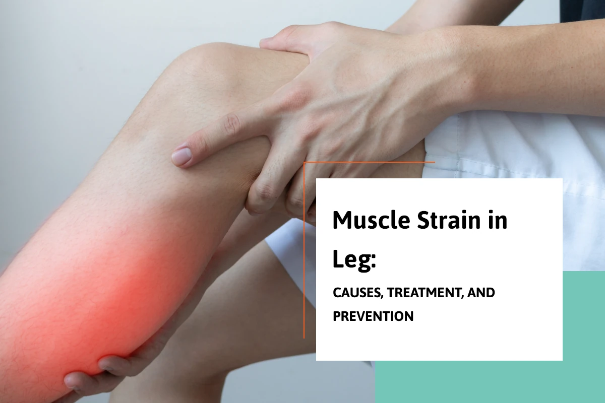
Muscle Strain in Leg: Causes, Treatment, and Prevention

Muscle Strain In Leg

A strained muscle in the leg can happen suddenly during physical activity or build up over time with repetitive use. Whether it’s a mild strain from jogging or a more serious injury while playing sports, knowing how to recognize, treat, and prevent muscle strains is critical for maintaining mobility and avoiding long-term issues. In this blog, we’ll discuss the risk of muscle strains, risk factors, how to reduce swelling, and effective treatments to promote healing and prevent future injuries.

What Happens When a Muscle Strain Occurs in Your Leg?

A muscle strain occurs when the muscle fibers are stretched beyond their limit or even torn. You might feel a pop or sudden sharp pain when the injury happens. This is often accompanied by immediate weakness in the injured area, making it difficult to continue activity. Sometimes, the strain is mild and causes minor discomfort, while other times it can be a complete muscle tear, known as a grade three strain.

Recognizing the Severity of a Muscle Strain In The Leg

Strains are typically classified into three grades:

  • Mild strain (Grade I): Minor damage to muscle fibers with little loss of strength or motion.
  • Moderate strain (Grade II): More significant injury with noticeable weakness, swelling, and restricted movement.
  • Severe strain (Grade III): Complete rupture of the muscle, often causing intense pain and the inability to use the affected leg.

In some cases, ongoing issues can lead to a chronic muscle strain, where the muscle has not fully healed and continues to cause discomfort during physical activities.
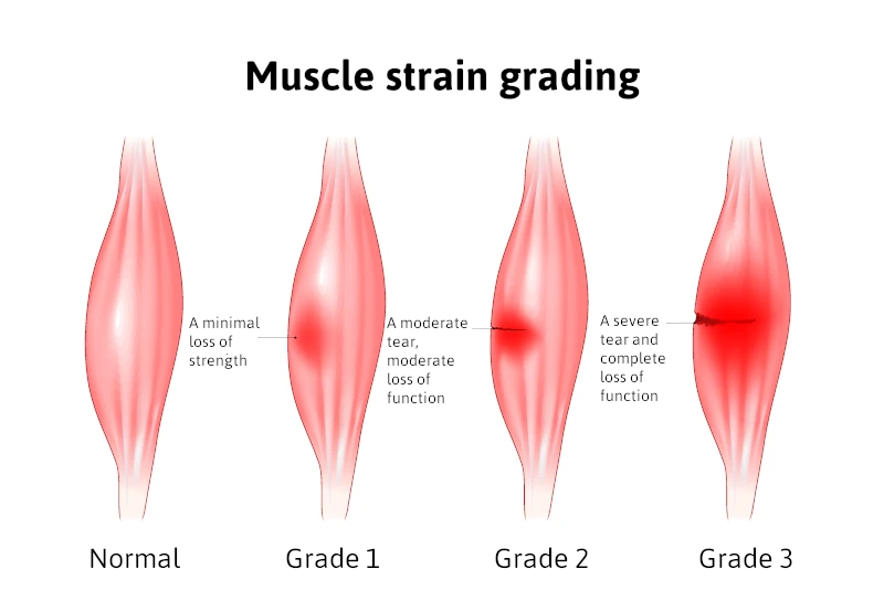
Grades of Muscle Strain In Leg Infographic

Risk Factors for Muscle Strains

Certain factors increase your risk of muscle strains, including:

  • Inadequate warm-up before exercise
  • Poor flexibility
  • Muscle fatigue
  • Previous muscle injuries
  • Age-related changes to muscle elasticity
  • Participation in high-intensity sports

Understanding these risk factors can help you take steps to protect yourself during activities.

How to Prevent a Muscle Strain in the Leg

Fortunately, you can take proactive steps to prevent muscle strains. Some of the best strategies include:

  • Proper warm-up and stretching before exercise
  • Strengthening exercises to support vulnerable muscles
  • Staying hydrated and maintaining electrolyte balance
  • Cross-training to avoid overuse of specific muscle groups
  • Gradually increasing the intensity of your workouts

Consistency in these practices can significantly lower the chance that a pulled muscle will sideline you.

Immediate Treatment: Rest, Ice, Compression, and Elevation

When a strain occurs, early treatment is crucial for minimizing damage and speeding recovery. Follow the Rest, Ice, Compression, and Elevation (RICE) method:

  • Rest the injured area immediately to prevent further muscle tearing.
  • Apply ice wrapped in a cloth for about twenty minutes every two to three hours to reduce swelling.
  • Use an elastic bandage for compression to limit inflammation.
  • Elevate the leg above heart level whenever possible to decrease swelling.

These steps are essential within the first 48 hours after the injury.

Medications That Can Help

To manage pain and inflammation, doctors often recommend non-steroidal anti-inflammatory drugs such as ibuprofen. These medications work to reduce both pain and swelling. However, it’s important to use anti-inflammatory medications as directed and not rely on them as a substitute for proper rest and rehabilitation.

Rehabilitation: Regaining Strength and Flexibility

After the initial swelling and pain subside, it’s important to begin gentle rehabilitation exercises. Restoring range of motion without causing additional injury is the goal. Activities might start with light stretching, followed by progressive strengthening exercises. Physical therapy is often beneficial, particularly for moderate to severe strains.

Ignoring a muscle strain or returning to activity too soon can prolong recovery and increase the risk of chronic muscle strain. Taking the time to fully heal ensures the best long-term outcomes.

When to See a Doctor for a Muscle Strain in Leg

Most mild to moderate strains heal well with home care, but there are times when professional medical attention is necessary. Seek help if you experience:

  • Severe pain and swelling
  • Inability to walk or bear weight
  • Numbness or tingling
  • Noticeable muscle deformity

These symptoms could indicate a grade three strain or another serious injury that might require more advanced interventions, such as imaging studies or even surgery.

Long-Term Outlook

With prompt and proper care, most people fully recover from a leg muscle strain and return to their normal activities. Severe cases may take several months, while mild strains often heal within a few weeks. Adhering to treatment recommendations and rehabilitation plans is essential for preventing a recurrence. Once healed, it’s wise to incorporate injury prevention strategies into your daily routine. This not only minimizes the risk of muscle strains in the future but also helps enhance overall performance during physical activity.

Conclusion

A strained muscle leg injury can be frustrating, but understanding the injury process and knowing how to respond makes a huge difference in recovery. Remember that the risk of strains is higher if you have certain risk factors, but proper preparation and caution can prevent muscle strains. Treat early with rest, ice, compression, and elevation, and support healing with anti-inflammatory medications like ibuprofen when appropriate. Always listen to your body — if you feel a pop or experience intense pain, take it seriously. Healing takes time, but with patience, you can fully recover and return to your favorite activities stronger than ever.

Schedule Your Comprehensive Foot Evaluation Today

Call us today at our Fort Collins location (970) 484-4620, Broomfield location (720) 259-5053 or use our online scheduling system to book your appointment.

With convenient clinical foot locations in Broomfield and Fort Collins, Colorado.
The Hidden Truth About Calcaneal Spur Surgery: What Your Doctor May Not Tell You

calcaneal spur surgery

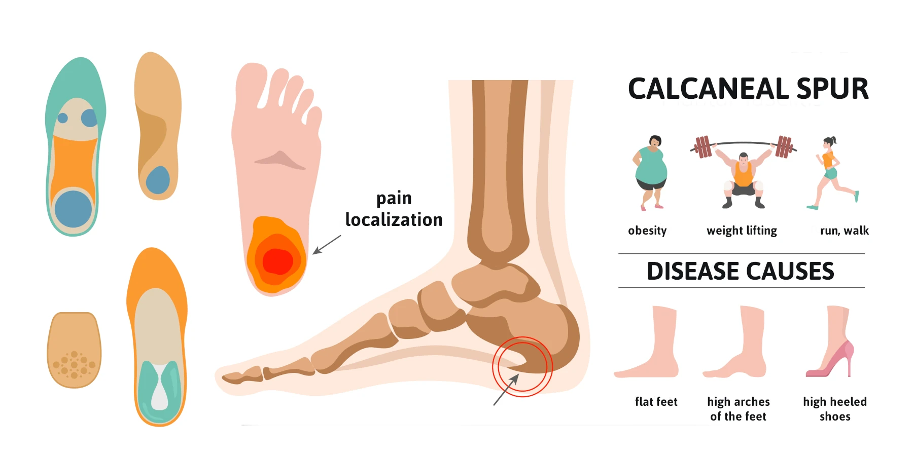
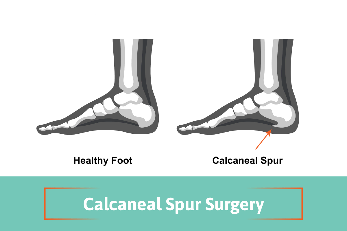
A calcaneal spur, often referred to as a heel spur, is a bony growth that develops on the heel bone (calcaneus). This condition is commonly associated with plantar fasciitis, which involves inflammation of the plantar fascia ligament, leading to significant heel pain. While many individuals manage heel pain through conservative treatments, some may require calcaneal spur surgery to alleviate persistent discomfort and restore mobility. This blog aims to provide a better understanding of why the removal of the heel spur is rarely needed and can be considered a fallacy. I will contrast the old-school ideas with the more recent concepts based on science.

What is a Calcaneal Spur?

A calcaneal bone spur is a bony growth that forms on the underside of the heel bone, typically at the site where the plantar fascia attaches to the calcaneus. Most believe that the point of the fascia from the heel bone at this location may stimulate new bone to form, thus causing the spur formation. In the 1930s, Dr. Spitz was the first doctor to suggest that the actual heel spur is not causing the pain, but rather the fascia that is pulling from the calcaneus.

This condition can arise due to repetitive stress and strain on the foot, often resulting from activities such as running, walking, or standing for long periods. The bone spur development can occur alongside plantar fasciitis, leading to significant heel pain and discomfort. Symptoms may include sharp pain in the heel, especially in the morning or after prolonged periods of sitting. While many individuals experience relief through non-surgical interventions, some may require surgery when conservative treatments fail to provide adequate relief.

Causes of Calcaneal Spurs

calcaneal spur surgery causes infographic

Calcaneal spurs commonly develop due to a combination of factors, including:

  • Repetitive Stress: Activities that involve excessive strain on the feet, such as running, dancing, or standing for extended periods, can lead to the formation of a heel spur.
  • Plantar Fasciitis vs Plantar Fasciosis: the old school concept is that the fascia is inflamed. This has been disproven by one study performed by Dr. Harvey Lamont. When he performed surgery to release the plantar fascia from the heel bone, specimens were sent to a lab. Out of 50 specimens, 49 showed fascial degeneration while only one showed inflammation. This supports that it is not an inflammatory condition.
  • Chronic pulling of the plantar fascia can contribute to the development of a calcaneal spur, as the body attempts to stabilize the area by forming new bone.
  • Foot Structure: Abnormalities in foot structure, such as flat feet or high arches, can increase the risk of developing heel spurs.
  • Age and Weight: Excess body weight can contribute to higher stress levels on the feet.

Symptoms of Calcaneal Spurs

Individuals with a calcaneal spur often experience a range of symptoms, including:

  • Heel Pain: The most common symptom is a sharp, stabbing pain in the heel, especially during the first steps in the morning or after prolonged sitting.
  • Swelling and Inflammation: The area around the heel may become swollen and tender to the touch due to inflammation.
  • Pain with Activity: Activities like walking, running, or standing for long periods can exacerbate heel pain.
  • Difficulty in Movement: In severe cases, individuals may find it challenging to walk or engage in physical activities due to discomfort. Understanding these symptoms is crucial for individuals to seek appropriate medical evaluation and treatment.

Diagnosis of Calcaneal Spurs

Diagnosing a calcaneal spur typically involves a thorough evaluation by a healthcare professional. The diagnostic process may include:

  • Medical History: The doctor will inquire about the patient’s symptoms, pain levels, and any previous foot injuries or conditions.
  • Physical Examination: A physical examination of the foot will help assess tenderness, swelling, and range of motion.
  • Imaging Tests: X-rays are often used to confirm the presence of a calcaneal spur and assess its size and location. In some cases, MRI or ultrasound may be utilized to evaluate soft tissue involvement, such as the plantar fascia and the Achilles tendon. An accurate diagnosis is essential for determining the most appropriate treatment options, whether conservative or surgical.

Non-Surgical Treatment Options

Before considering surgery, healthcare providers typically recommend conservative treatment options for calcaneal spurs, including:

  • Rest and Activity Modification: Reducing high-impact activities and allowing the foot to heal can alleviate symptoms.
  • Physical Therapy: A physical therapist can design a personalized program that includes stretching and strengthening exercises for the foot and ankle, improving flexibility and reducing pain.
  • Orthotic Devices: Custom orthotics or shoe inserts can provide additional support and cushioning to the foot, helping to redistribute pressure away from the heel.
  • Pain Medication: Over-the-counter medications, such as ibuprofen or acetaminophen, can help manage pain and inflammation.
  • Cortisone injection vs Regenerative medicine: Corticosteroid injections may be helpful but rarely provide permanent relief and this is because the fascia is not inflamed, but rather degenerating based upon studies. Therefore, the option of regenerative medicine provides a very success rate to reverse the symptoms on a long-term basis as this treatment helps repair the fascia that is degenerating.

When is Surgery Necessary?

Surgery for calcaneal spurs is generally considered when conservative treatments fail to alleviate pain and improve mobility after several months. Indications for surgery may include:

  • Persistent Pain: Severe, ongoing heel pain that significantly affects daily activities and quality of life.
  • Diagnosis Confirmation: Confirmation of a calcaneal spur through imaging tests and persistent symptoms despite conservative management.
  • Functional Limitations: Difficulty participating in work, recreational activities, or exercise due to heel pain. Surgical intervention aims to remove the spur and address any accompanying issues, such as plantar fasciitis, to restore function and reduce pain.
  • Regenerative Medicine: The option of regenerative medicine has greatly reduced the need for surgery. (see testimonial videos)

The Surgical Procedures for a Calcaneal Spur

Contrasting heel spur surgery to endoscopic plantar surgery. Calcaneal heel spur surgery usually involves incision on the side of the heel and the spur is removed along with the attachment of the fascia to the heel bone at the spur location. This is a procedure that surgeons have not performed since 1990. Because the heel spur is not thought to be the cause of your pain, using a small incision to place a scope across the bottom of the heel allows the surgeon to cut a portion of the fascia so that it may stretch and elongate and reduce its pull on the heel bone. This technique has been adopted by majority of the Podiatric Foot and Ankle Surgeons, and because of its high success rates, it supports that fact that the heel spur is not the cause of your pain.

Recovery from heel spur surgery

This type of surgery usually involves a larger incision and more trauma and longer recovery with immobilization in a cast, and sometimes non-weight bearing.

Recovery from endoscopic heel surgery

This type of surgery uses a small incision, and you can bear weight immediately and may return to wearing shoes in 3-4 weeks.

Watch Patient Testimonial Videos


Post-surgery, patients are typically advised to avoid weight-bearing activities for a specified period to allow for proper healing. Physical therapy may be recommended to facilitate full recovery and regain strength and range of motion.

Recovery and Rehabilitation for Calcaneal Spur Surgery

The recovery time for calcaneal spur surgery varies among individuals, with most patients experiencing significant improvement within a few weeks. A typical recovery process includes:

  • Pain Management: Pain may be managed with prescribed medications and the application of ice to reduce swelling.
  • Physical Therapy: Engaging in physical therapy is crucial for restoring strength, flexibility, and balance. Therapists may focus on exercises to improve range of motion and stability, particularly around the Achilles tendon and plantar fascia.
  • Gradual Return to Activity: Patients are typically encouraged to gradually return to normal activities as healing progresses, following their surgeon’s recommendations.
  • Long-Term Management: Maintaining a healthy weight, wearing appropriate footwear, and incorporating regular stretching and strengthening exercises can help prevent future heel issues.

Conclusion

Calcaneal spur surgery can provide significant relief for individuals suffering from persistent heel pain due to calcaneal spurs. While many patients find success with conservative treatments, surgery remains an effective option for those who do not respond to traditional methods. Understanding the causes, symptoms, and treatment options for calcaneal spurs is essential for making informed decisions about care. If you suspect you have a calcaneal spur or are experiencing persistent heel pain, consult with a healthcare professional to explore the best treatment options for your needs.

Lateral Foot Pain Slowing You Down. Finding The Source!

lateral foot pain

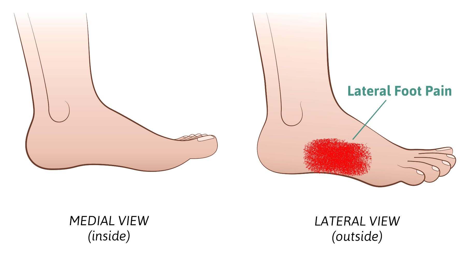
Lateral foot pain refers to the pain being on the outer side of the foot. This means anywhere from the small toe joint back to the heel bone area. Any pain that is along the outer edges of the foot. This blog will review many problems that can arise in this region of the foot. However, before we get started it’s important to point out that lateral foot pain in many cases may be related to a problem elsewhere in the foot. This other issue is creating a situation where you have difficulty walking and favor the painful area by rolling your foot towards the outside. This may create stiffness and pain in the lateral foot. So as a foot specialist I’m always looking to find in the patient’s story, their history, was there some other type of pain that preceded the lateral foot pain. Here are six things that cause lateral foot pain and treatment options.

  • Cuboid syndrome – This pain is usually associated with the calcaneal cuboid joint. This joint lies on the outside of the foot at the highest part of your arch on the lateral side of your foot. The history of plantar fasciitis is commonly associated with cuboid pain. By favoring the foot and walking on the outside of the foot the reducing the pulling of the plantar fascia from the bottom of the foot and thus significant stress is placed in this area. AS the heel pain is reduced the cuboid pain increased. Treatment therefore should start by addressing and eliminating the heel pain and the cuboid pain will fix itself.
  • Peroneal tendonitis – The peroneal tendons lie on the outside of the foot coming from behind the fibula bone and then coursing on the outside of the heel One these tendons, the peroneus brevis’s attaches to the base of the fifth metatarsal, the large eminence that protrudes halfway between the ball of your foot and the heel. The other tendon, peroneus longus runs beneath the arch and attaches to the base of the first metatarsal bone and helps to pull this bone downward to support the medial arch. Basically, both tendons are like stirrups helping to stabilize the foot from moving side to side. Any activity that requires along side-to-side motion can stress these tendons and, in some situations, they can develop tears. Flat feet may also contribute to peroneal tendonitis. We have found that human cellular tissue products are a great option to help repair these tendons so surgery can be avoided.
  • Fractures – Fractures of the heel bone cuboid bone or fifth metatarsal can all cause pain in this area. Of these three fifth metatarsal bone fractures are the most common.
  • Fifth metatarsal bone fractures – these are usually associated with an ankle sprain where the twist is so forceful the peroneal brevis bone that attaches to the base of the fifth metatarsal pulls a portion of the fifth metatarsal away from the rest of the bone. This is referred to as an avulsion fracture. The other type of fracture referred to as a Jones fracture occurs when upward force is placed primarily on the fifth metatarsal and not the other metatarsals causing the bone to fracture just behind the base of the bone. An example would be a basketball player landing on another player’s foot with all the body weight on the fifth metatarsal. Both these fractures require immobilization in a cast or Cam-boot and the probability of surgery to repair the fracture is more common with the Jones fracture.
  • Cuboid fractures – This bone is between the heel bone and the fifth metatarsal. Fractures here are very uncommon and may be associated with trauma but more likely may be caused from overuse.

Treatment for lateral foot pain is usually immobilization

  • Calcaneal fractures – The calcaneus is your heel bone and fractures can occur from trauma such as a fall and when severe may require surgery. However, in most cases overuse can cause stress fractures the heel. The heel bone is very vascular, so the bone tends to heal very well.
  • Sinus tarsi syndrome – This is very common and is associated with the subtalor joint. This joint lies beneath the ankle joint and is the joint that allows your foot to move like a universal joint on a care. In all different body planes. There are two ligaments in the joint that are thought to become inflamed from overuse or trauma such as an ankle sprain. It’s not uncommon for someone to report an ankle strain weeks or months ago but the pain may linger in this location. Treatment with cortisone injections and custom-made arch supports called orthotics successfully eliminates the pain.
  • Sural nerve pain – This is a nerve that originates in the lower leg and travels down the side of the foot to the small toe. This nerve can be irritated. By a multitude of different types of traumas including, Surgery with an incision being placed on the outside of the foot or ankle. From an ankle strain causing the nerve to be stretched. Surgery or trauma such as being kicked in the lower leg. Treatment may include, cortisone injections, laser treatments and when pain is severe and conservative measures fail, surgery removal on the nerve in the leg and burying into muscle to prevent regrowth.
  • Great toe joint pain – This requires its own special category as any pain in the great toe joint area will cause you to walk differently. Because normal ambulation requires a significant amount of body weight to be transferred onto the great toe joint, anything that alters the normal gait pattern can cause significant lateral foot pain. This would include the following
  • Bunion pain – this occurs when the great toe angulates towards the second toe and a bump is created at the base of the great toe joint. Shoes will cause irritation to the bump.
  • Sesamoiditis – There are two sesamoid bones the sit beneath the great toe joint and function like the kneecap to improve the strength of the muscles that pull your great toe down. This can become painful for many reasons. Again, you’ll tend to reduce the load on these bones and transfer your weight laterally.
  • Hallux rigidus – Hallux refers to the great toe and rigidus refers to great toe joint having limited motion. When the great toe cannot move up adequately for normal gait you’ll have to transfer weight to the outside of your foot. Treatment for all three of these conditions may include custom made arch supports, orthotics, to reduce pain and improve function. Cortisone injections. Human cellular tissue products to reduce joint pain for Hallux rigidus, see our video below. Or surgery is another way to correct the problem.

Treatments for lateral foot pain

  • In the early stages taking anti-inflammatories and physical therapy may help reduce swelling along with an elastic bandage early on.
  • Reduction of physical activity may also be useful.
  • Use of shoe inserts may be useful in a treatment plan..

Final thoughts. So, if you’ve been dealing with lateral foot pain and are not sure what it is and what treatment choices you must consider consulting with a foot and ankle specialist. Finally remember you could be wasting a lot of time and money treating lateral foot pain that is failing because it never was the primary problem. Give us a call at one of our local foot and ankle clinics in Fort Collins and Broomfield. We see patients searching for ‘the best foot doctor’ from all across the tri-state area, such as Wyoming, Cheyenne and Loveland. With over 10600+ views on Youtube and a large Facebook community, we are always looking to provide real solutions for all our patients foot ailments.

Call us today at our Fort Collins location (970) 484-4620, Broomfield location (720) 259-5053 or use our online scheduling system to book your appointment.

Tibialis Anterior Tendonitis: Symptoms and Treatment Options

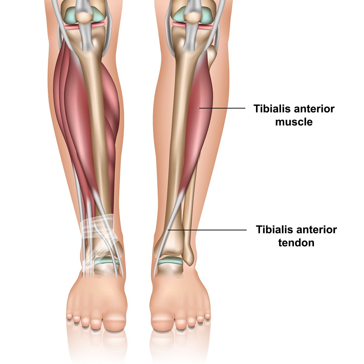
Muscles and tendons tibialis anterior tendonitis

The foot has several tendons that originate in the lower leg and extend into the foot. They each have different functions to stabilize the foot and help us to propel our bodies forward when we walk or run. The anterior tibial tendon is one of these tendons. Although tibialis anterior tendonitis may be the least common complaint of those experiencing tendon pain around the ankle joint, it’s important to understand how to recognize and what can be done to eliminate your pain.

Anatomy and function- The anterior tibial tendon is the third largest tendon in the foot and ankle. The largest is the Achilles tendon and the second largest is the posterior tibial tendon. The anterior tibial tendon originates as a muscle in the lower leg adjacent to the shin, tibia, and extends down to the foot. It is on the front of the ankle. It inserts onto the top and inner side of the arch.

Its function is to pull the foot up and support the arch. It also assists in supinating the foot. Supination is a motion often referred to and means that the arch of the foot increases and the foot angles towards the opposite foot. Finally, it decelerates the downward motion of your foot towards the ground. From the time your heel contacts the ground to the point of the ball of the foot bearing weight it prevents your foot from slapping against the ground. So, it’s very important for normal gait to occur. It helps to identify the tendon by pulling your foot up towards you and it will be the tendon that protrudes on the front of the ankle and is running towards the inside of the foot towards the big toe.

Tibialis anterior tendonitis is a condition that causes pain and discomfort along the front of the lower leg and ankle due to inflammation or degeneration of the tibialis anterior tendon. This tendon plays a crucial role in foot movement, particularly in lifting the foot upward while walking or running.

Where Will The Pain Be If I Have Tibialis Tendonitis?

It is most common towards the front of the ankle area. The symptoms of tibialis anterior tendonitis may also create pain above the ankle in the lower leg adjacent to the tibia. This may be referred to as anterior shin splints. Referring to shin splints on the front of the leg. Symptoms may also be closer to or at the point where the tendon attaches to the foot on the inner side of the arch.

What Causes Tibialis Anterior Tendonitis?

Several factors can contribute to tibialis anterior tendonitis, including:

  • Overuse – this may occur from doing too much too soon which can happen in any activity. However, any activity that requires you to extend your foot in front of the knee joint may increase the probability of overuse. Examples of this may be racquet sports, hiking, or running downhill.
  • Biomechanics – If your born with a flat foot the tendon may have to work harder to help support your arch.
  • Improper running technique – Over striding while running can also cause overuse of the tendon. By taking shorter strides you won’t be extending the foot in front of you as much as this will help to reduce the need for the anterior tibial tendon to stop foot slap.
  • Neurological problems – Neurological problems may not cause pain along the tendon, however, may cause damage to the nerves that stimulate the anterior muscle to contract. This is referred to as drop foot. Drop foot can be associated with back issues of a nerve issue around the see. Nerve surgery called decompression surgery can help with this.
  • Trauma – a severe ankle sprain could cause a tear to the tendon. Also, a laceration to the ankle area may also cause a partial or full tear.
  • Shoe gear – High heels may also be a cause and they will put more demand on the tendon to reduce the potential for foot slap because of the elevation of the heel off the ground.
  • Improper Footwear: Wearing unsupportive or worn-out shoes can alter foot mechanics, leading to excessive strain.

Symptoms of Tibialis Anterior Tendonitis

Common signs and symptoms of tibialis anterior tendonitis include:

  • Pain and tenderness along the front of the shin, ankle, or top of the foot.
  • Swelling in the affected area, especially near the tendon insertion.
  • Weakness or difficulty lifting the foot, potentially leading to a “foot slap” while walking.
  • Stiffness and discomfort that worsens with activity, particularly when walking downhill or running.
  • Pain when pressing on the tendon or flexing the foot upward against resistance.

Diagnosis or Evaluation of Tibialis Anterior Tendonitis

A podiatrist will typically diagnose tibialis anterior tendonitis through:

  • Clinical Examination: The physical examination alone will give the doctor a good idea if the tendon is damaged. There will be pain and swelling along the tendon or its muscle higher up the leg. Also, weakness of the tendon may be noted.
  • X-rays – these will not show tendon damage but are useful to rule out other problems.
  • Diagnostic Ultrasound: Assessing the tendon’s structure and detecting any tears or degeneration.
  • MRI Scan: MRI scans or diagnostic ultrasound are useful to confirm the diagnosis and see the extent of the tendon damage. The diagnosis will frequently be tibialis anterior tendinopathy. The tendon may be damaged but not torn.
  • Gait Analysis: Observing walking patterns to detect abnormalities such as foot drop or imbalance.

Treatments for Tibialis Anterior Tendonitis

Most cases of tibialis anterior tendonitis can be managed with non-surgical treatments, including:

  • Rest and Activity Modification: Reducing strain by limiting high-impact activities.
  • Cold Therapy: Applying ice packs to reduce inflammation and pain.
  • Anti-inflammatory Medications: Nonsteroidal anti-inflammatory drugs (NSAIDs) may help relieve symptoms.
  • Immobilization: Wearing a CAM walker or brace to reduce movement and promote healing.
  • Orthotic Devices: Custom arch supports to help alleviate stress on the tendon.
  • Physical Therapy: Stretching, strengthening exercises, and modalities like ultrasound therapy can aid recovery.

Advanced and Surgical Treatments

For severe cases where conservative measures are ineffective, the following treatments may be recommended:

  • Laser Therapy: Multiwave laser (MLS laser) MLS treatments have shown success in reducing pain and inflammation.
  • Human Cellular Tissue Products: Also known as stem cell therapy, these injections can promote tendon healing. This treatment has significantly reduced the chance of surgery when partial tears are noted. It is important to note that in many cases the tendon may not be inflamed as much as it is breaking down. This is sometimes referred to as tendinosis rather than tendonitis. For this reason, regenerative medicine is very appropriate as it helps to repair the tendon.
  • Corticosteroid Injections (With Caution): While cortisone injections may provide short-term relief, excessive use can weaken the tendon and increase the risk of rupture.
  • Surgical Repair: If a partial or full tear is noted surgery to repair the damage may be recommended. With a full tear weeks of non-weight bearing may be needed. In cases of complete tendon rupture or severe degeneration, surgical intervention may be necessary to restore function. Post-surgery, a period of non-weight-bearing followed by rehabilitation is typically required.

How to Avoid Tibialis Anterior Tendonitis?

It’s important when taking up a new activity to not do too much too soon. Also, if you suffer from flat feet, make sure you have supportive shoes or consider over-the-counter arch supports or custom-made orthotics.

Hopefully this blog will help you to better understand if you have tibialis anterior tendonitis and how some of the newer treatments available including regenerative medicine and laser treatments can help manage the condition. Our foot and ankle clinics cater to patients from Colorado, Wyoming and Nebraska. We also see patients from all over the United States and even our neighbors from Canada who have searched for a ‘expert podiatrist near me‘. Watch our patient testimonials on YouTube as we now have over +11000 followers.

For expert evaluation and treatment, schedule an appointment with Anderson Podiatry Center today.

Call us today at our Fort Collins location (970) 484-4620, Broomfield location (720) 259-5053 or use our online scheduling system to book your appointment.

Say Goodbye to Foot Pain: Effective Cuboid Syndrome Treatment

Cuboid Syndrome treatment

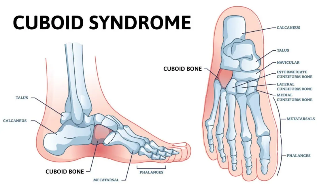
What is Cuboid Pain? Cuboid pain is not one of the most common foot complaints, but that’s why this blog is important for you to read. It’s a diagnosis that can be overlooked by healthcare professionals. The cuboid bone is a cube-shaped bone on the outside, lateral aspect of our foot – the pinky toe side of the foot. It lies halfway between our toes and heel. If you were to look at your foot from the side when you’re not bearing weight, it would be the bone that is at the highest part of your arch.

This area is called the lateral arch. The lateral arch is formed by the 4th and 5th metatarsal bones, which are the long bones that extend out to the ball of the foot that your bear weight is on. These two bones are connected to the cuboid bone by ligaments, and the bone forms the joint on the other side of the cuboid bone. The medial arch is the inner portion of the arch and is formed by the three metatarsal bones that connect to the midfoot. The medial arch is thought to be stronger than the lateral arch and can take more stress placed through this portion of the foot. This basic understanding is important to know as it plays a role in why you get cuboid pain. We will discuss the symptoms, diagnosis and cuboid syndrome treatment options in this blog.

What Happens When you Have Cuboid Syndrome Pain?

It is thought that the cuboid bone gets out of proper alignment with the calcaneus (heel) bone. This misalignment of the cuboid bone will cause pain in and around the joint. It can also be described as a midfoot sprain. It may also be referred to as cuboid subluxation.

Risk Factors for Cuboid Syndrome Include:

  • Trauma– ankle sprains, often described as an inversion ankle sprain. When this occurs, the ankle twists in such a way that the body weight rolls towards the outer side of the foot on the small toes’ joint side. The added weight in this area can cause displacement of the calcaneocuboid joint. Other types of foot injuries may also be a cause.
  • Foot biomechanics – individuals who overpronate or flatten their feet too much may be more prone to cuboid syndrome. It is thought that overpronation is the cause of cuboid pain for 80 percent of the population.
  • Repetitive stress – overdoing it, especially with weight-bearing activities, can also lead to this problem. Ballet dancers are especially prone to this injury because of the positions the activity requires their feet to be placed in.
  • Compensatory pain – one of the more common causes can be from pain elsewhere in the foot or ankle. We are very good at modifying our gait to help prevent pain. After a while, we may not even be aware we are doing it. If another foot problem requires us to walk on the outside of the foot to make it feel better, this may also be a cause. One example of this is plantar fasciitis. This will cause pain in the bottom of the heel, and it is common to compensate and avoid this pain by rolling your foot to the outside. If this is the case, it’s very important to resolve this pain first, and in many cases, this can fix the cuboid pain once the compensation stops.

What will my Symptoms be?

There will be pain on the outer side of the foot midway between the heel and the ball of the foot. The pain will occur when putting weight on the foot. It can be very severe and is usually a dull, deep pain. You may find you feel better when walking in shoes than barefoot as the support of the shoes may help with the discomfort.

How will the Doctor Diagnose Cuboid Syndrome?

The doctor can learn a lot from your explanation of where the pain is. Also, examination of the foot will be important as the pain will tend to be directly over the calcaneocuboid joint. There may also be some swelling. X-rays will also be taken to rule out fractures of the cuboid or adjacent bones. Other problems that could mimic cuboid syndrome include a fracture of the cuboid bone, tendonitis of tendons in the area called peroneal tendons. MRI or diagnostic ultrasound may be used to evaluate these other problems to determine what cuboid syndrome treatment will be the most effective.

Mysterious Pain on the Outside of the Foot? Here are the Treatment Options for Cuboid Syndrome!

  • Physical therapy – A physical therapist may be able to reduce pain in the cuboid area. This can also help to prevent the recurrence of cuboid syndrome.

This can be accomplished with

    • Balance exercises – By improving balance, you will be less likely to injure your foot as ankle sprains can be a common cause of cuboid syndrome.
    • Mobility exercises – To help regain full range of motion and get rid of any stiffness that may have occurred with cuboid syndrome.
    • Strengthening exercises – this will provide more strength to support the foot and ankle. Stronger muscles and tendons will provide more support to the foot and ankle and reduce the chance of cuboid syndrome.
    • Support of the cuboid bone – This may be accomplished by using padding underneath the cuboid bone area, over-the-counter arch supports may also help.
  • Orthotics – Custom-made inserts to go in your shoes may be the most important part of the treatment that your podiatrist can provide. This can give very precise support to that portion of your foot and lead to full recovery. It must be emphasized that orthotics may be used to treat the primary problem that created the cuboid syndrome. A very common example would be if you’ve been favoring your heel (plantar fasciitis) and this compensation has put added stress on the cuboid area. Walking differently and favoring your heel makes it feel better, but now you have cuboid pain. So by eliminating the heel pain with orthotics, the cuboid pain goes away.
  • Taping the foot and ankle – Taping the foot may also help support the calcaneocuboid joint area. This is especially effective in the early stage of the problem.
  • RICE – Rest, ice, compression, and elevation may also be used. These treatments will reduce inflammation in the area and promote healing. For some, resting the foot for up to 6-8 weeks may be necessary before full recovery and returning to regular activities.
  • Manipulation – This should only be attempted by a healthcare professional familiar with this technique. It is sometimes referred to as the cuboid whip. The technique involves placing the patient on their stomach with the knee on the involved side flexed. Then, with both hands gripping the foot, it is brought upwards and then thrust rapidly downward. This will allow the cuboid to pop back into its proper position.
  • Modifying activities– Cross-training may also be helpful. If you’ve been doing weight-bearing activities that involve a lot of side-to-side motion which may stress the cuboid joint area more, you may elect to take a break and bike,

As with many foot and ankle injuries, cuboid syndrome should be properly diagnosed and treated as soon as possible. This will help to speed up getting back to full activities. So, if you’re having a mysterious pain on the outside of your foot please consider the possibility of cuboid syndrome. We have skilled podiatrist who guide each of our patients to ensure a proper cuboid syndrome treatment is chosen that best suits the individual.

Call us today at our Fort Collins location (970) 484-4620, Broomfield location (720) 259-5053 or use our online scheduling system to book your appointment.

What to Know About Arthritis Foot Care and What You Can Do

Arthritis is a very common problem in the foot and ankle area and can occur in different locations. Because the foot has so many bones, there are many joints that can become painful. 

arthritis in feet causes, symptoms, and treatment

Symptoms

First, it is important to understand what the symptoms of arthritis are. Symptoms tend to present as a deep, dull type of pain that occurs when you first get up in the morning, Then after you take a few steps, it can gradually get better.  As the arthritis becomes more severe, it can present all the time, particularly when you’re active. Understand that arthritis occurs when the cartilage that lines the joint becomes worn down and thinner. In severe situations it can wear down to bone.  Cartilage is very slick- even slicker than ice! It acts that way to let your joints move more easily.   

Causes

There are a multitude of causes that can result in arthritis. It could be from gout attacks, where inflammation in the joints destroys the cartilage, or even rheumatoid arthritis, where patients have autoimmune diseases that can affect the joints.

There may have even been an injury to the joint or bone that caused the cartilage to be damaged. This is especially common in the ankle joint in the case of someone who is a chronic ankle sprainer or has had an ankle fracture.  All these conditions can be called “secondary arthritis” as they are related to another issue. 

For many that suffer from arthritis, it is commonly referred to as primary arthritis, meaning there is no direct cause. As we age from using our joints, the cartilage tends to wear down

Lastly, the most overlooked cause is foot structure. Very few doctors are trained  to evaluate foot structure and foot biomechanics. Some foot types, particularly flat feet, might lend themselves to wearing down joints more than feet that have a normal biomechanical structure.

 Where does arthritis occur most commonly?

 It occurs most commonly in the great toe joint, ankle and midfoot. In the great toe, it will frequently cause a stiff toe joint, referred to as Hallux Rigidus.  It can also occur in the midfoot area, halfway between the toes and the ankle in the arch area. This is also more common as we get older. Finally, it can occur in the ankle joint, especially if you have had multiple ankle sprains, or if you have any kind of history of ankle fracture- this lends itself to a greater possibility of having ankle arthritis at some point in your life.

Five things you can do

  • Avoid inflammatory foods: Foods high in sugars and carbohydrates should be reduced. Why? Because they create inflammation in your body. Breads, sugar and white flour should be avoided. Also watch out for fructose as this is used in many beverages such as fruit juices and is probably worse than just scarfing down a candy bar. Instead of fruit juice, choose to eat the actual fruit as it’s much healthier for you. This will also help keep your weight down.    
  • Exercise: Yes, that is right, keep moving. Maybe if you used to run and it bothers you too much, consider biking or walking. You may want to take on swimming or other activities. Make sure to modify the frequency and duration because motion helps your body create more growth hormones which can help repair your joints. 
  • Consider supplements: Supplements such as Turmeric, Sam-e , Hyaluronic Acid and Collagen may also be effective and help fight inflammation. 
  • Avoid anti-inflammatories: While they may make you feel good today, the long-term effects are negative for your joint health. An exception to this would be if you have an autoimmune disease such as rheumatoid arthritis that is managed by your doctor. 
  • Use more supportive shoes: If you think you may be an overpronator, meaning you have flat feet, supportive shoes should be considered. You may also want to use an inexpensive over the counter arch supports. 

Treatment

  • Support the foot and ankle: Oftentimes orthotics are recommended as they are custom-made inserts that go in the shoes. They are especially important to help stabilize the bony areas that can lend themselves to arthritis.
  • Repair and restore the joint: At Anderson Podiatry Center, we do this using stemcell treatment. Regenerative medicine is something we have been using now for over a decade and has been extremely useful for ankle joint problems, great toe joint pain, and pain in the midfoot area and the mid arch. It has proven to show improvement 80-90% of the time. 
  • Block your ability to feel the pain: We do this by shutting off pain signals to the brain.   In some cases, especially if the arthritis is in the midfoot or up by the ankle, we can remove one or two branches in the lower leg that block your ability to feel the arthritic pain in your foot or ankle.  This is also especially useful and has prevented many patients from going through ankle replacement surgery or ankle fusion surgeries (which have a significant risk factor and recovery time). 
  • Joint surgery: In the worst-case scenarios, when all else fails, there may be a need for revision of a joint by scoping it in the example of an ankle joint. Or finally a joint replacement or fusion. 

In summary, if you have these problems, it is important to understand that there’s a lot that you can do to repair your condition. And at Anderson Podiatry Center, we can offer non-surgical approaches and low invasive procedures such as stem cell or nerve resection, which may avoid more major joint replacement surgeries. Do not assume that you have to have an ankle joint replacement or other joints fused to treat your arthritis. Consider the options that you can do first, and if you are still struggling and want to get back to full activity, consider our unique approach. 

Sports and Exercise Training – Ouch! What do I do if I’m Injured?

Sports and Exercise Training – Ouch! What do I do if I’m Injured?

By Anderson Podiatry Center, in consult with Dr. Stephen Wilkinson, DPM, Sports Medicine Specialist

Congratulations! You’re training for an event, have begun or are maintaining an exercise routine and are motivated to compete or sustain your regime habit. You are to be applauded for taking your fitness and health seriously and incorporating it into your lifestyle. Your happy body, mind, work and social life will reward you for your efforts in a big way!

At some point in your fitness life, however, you will very likely experience pain or an injury that will frustrate or sideline you. Do you keep going? Do you take time off? As a sports medicine doctor, Dr. Wilkinson hears this a lot and has experienced it himself. How you manage these injuries, minor or not, is critical in determining if you can make your event goal or keep your downtime to a minimum and maintain your active life.

In his practice, at Anderson Podiatry Center for Regenerative Medicine, Dr. Wilkinson counsels many people to recover from injuries as quickly as possible and approaches his patient’s care and training plan with these steps and processes in mind, which helps assure they will be up and at it without skipping much of a beat. In this first of a series, we’ll present the initial steps that need to be taken to obtain a complete analysis that forms the basis of a sound and effective treatment and training plan. Then, later on, we’ll go into some of the customized specific treatment options and interventions – physical, mental and nutritional – that are utilized to get you to the finish line fast!

Step One:

A Complete evaluation begins with understanding the injury. An in-depth history is used to isolate the problem by asking questions about

  • The nature, location, and duration of the injury
  • The onset, course, and aggravating factors
  • Any treatments tried thus far

Patients often receive advice via their peer’s experiences or from the internet without a complete understanding of how that advice affects them specifically. Such treatment or training advice can unwittingly lead to injury. Patients often continue these improper training methods beyond the point of injury and then require professional help before they can continue towards their goals. We’ll alleviate this problem with a careful and complete history of the injury.

Step Two:

Step two involves taking time to understand what the patient is trying to accomplish in detail
(weight loss, cardiovascular fitness, a race completion, a time improvement etc.).  

  • Intervention is different depending upon whether the desired result is subjective or objective.

This breaks down to two basic groups of people:

  1. This group is seeking overall fitness and use a specific goal to provide motivation for training. They want to reach the finish line regardless of how long it takes. This requires significant core and mental fitness.
  2. This group has established overall fitness and is pushing to improve their fitness level via improvement in time or distance; more ‘fine tuning’ and achievement of maximum capacity
    while avoiding injury.

Step Three:

Step three involves understanding the patients starting point and whether or not his or her goal is reasonable to obtain by the time the patient needs to be ready to complete.

  • A 10% improvement per week is our target.

In this step, distance, intensity or speed of training are advanced 10% per week. Patients can often become impatient without clear guidelines and try and accelerate their training
faster than this tried and true path resulting in injury and training setbacks. “Slow and steady wins the race” as they say. Good advice to heed! The steps above give us a great foundation from which to build a plan of action to get you recovered and on your way. In the next blog we’ll discuss the nutritional, physical and mental approaches that provide the answers you need to get past your injury and help prevent future occurrences. See you then!


If you have an important goal to accomplish and are hindered by running or exercise pain, foot problems or injury, ankle injuries, plantar fasciitis or other problems, give me a call at
Anderson Center for Regenerative Medicine. I am not only ready to listen and understand, I am also ready to help.

To make an appointment with Dr. Wilkinson, Click HERE

Dr. Wilkinson is an ultra-runner, having completed many 50 and 100-mile races. As a Podiatrist who has successfully weathered his own sports injuries in the past, Dr. Wilkinson enjoys working with patients who are also athletes pursuing their own fitness goals. He believes in injury prevention through gait analysis, biomechanics, and sound, reasonable training techniques but is also an astute provider of clinical and surgical methodology to help return injured athletes to their playing field. Learn more about sports medicine injury treatment

Sports and Exercise Training – Ouch! What do I do if I’m Injured? Part 2

Sports and Exercise Training – Ouch! What do I do if I’m Injured? Part Two of Two

By Anderson Podiatry Center, in consult with Dr. Stephen Wilkinson, DPM, Sports Medicine Specialist at Anderson Podiatry Center

Injury Treatment Plan
In our last blog we covered the steps Dr Wilkinson takes to obtain a complete analysis that forms the basis of a sound and effective treatment plan for foot and ankle and lower extremity injuries. Those who become injured during training can benefit from an efficient, effective treatment plan to keep downtime to a minimum and still be able to reach their fitness or event goals.

This time we’ll explore some of the nutritional, physical and mental approaches Dr Wilkinson uses to help you get past your injury, get to your event or goal and help prevent future occurrences.

At Anderson Podiatry Center and our Center for Regenerative Medicine, Dr Wilkinson uses a mind and body restorative process and training redirection to help you meet your goals. This includes addressing nutrition, sleep, mental attitude, injury mechanism, training or exercise schedule as well as equipment and the potential of your current injury. He thereby devises a rehabilitation program specific to your needs. Advanced modalities are used when needed to accelerate healing. The following explains his approach to these individual parts of the rehabilitation process:

Moving Past your Injury – Focus On:

1. Nutrition

  • Although a basic nutrient allocation of 20-30% fat, 10-30% protein, and 45-65% carbs is optimal for those with an active lifestyle, requirements of activity, work and family make this easier said than done. Dietary balance should also be adjusted based upon the unique situation of each athlete. I.e. heavier athletes may include weight loss as a goal, and injured athletes may benefit from an adjustment in the recommended nutritional ratios and/or additional supplements.
  • It is not uncommon to see deficits in calcium, magnesium, vitamin D and iron. Test values are used to indicate replacement or supplements in the correct combination.

2. Sleep

  • The demands of modern life have contributed to a Healthy People.gov report that approximately 25% of average Americans have insufficient sleep half of the time. This, unfortunately, includes athletes, who have increased sleep demands during training, injury or illness. Sleep evaluation and intervention often involves a sincere allotment of time per daily and required activities, in addition to assessment with regards to activity required for sports goal success. Lower priority activities may need to be curtailed: a potentially tough decision but something’s gotta give to provide the long-term sleep requirements for progress in training and/or injury healing.
  • When sleep requirements for training exceeds the amount of daily sleep time available, additional time may be needed to safely reach goals despite many athletes’ desires to charge forward. Here, the vision and experience of a sports medicine consultant can be essential.

3. Mental Attitude

  • Possibly the most beneficial development that occurs when we strive to achieve more involved fitness or event goals is the process through which athletes improve their mental attitude. Improvement in self-confidence and improved self-image can often elevate an athlete’s success capacity beyond what was formerly believed. Important in this process is the conscious control of physical and emotional stress which must include appropriate and attainable goals and small interval reward breaks to allow for physical healing and mental reward for a job well done.
  • Visualization and self-talk, either alone or with a motivation/visualization coach is important preparation for success. Mental preparation and mental toughness require the acceptance of a fluid process with ups and downs in reaching a goal. The ultimate success of this process involves arriving at the final event both physically and mentally prepared.

4. Injury Mechanism

In foot, ankle and lower extremity sports injuries, understanding the injury mechanism is critical to successful intervention. In sports injuries, even in the most complex cases, there is typically a single event or pattern of recurring events that is at fault.

  • Single act injuries are often improved and resolved by isolation, rest, and regeneration.
  • Injuries from chronic repetitive mechanisms can often be overcome through improved balance and redirection of forces to other targeted areas.

The sports medicine consultant can be essential in determining the offending mechanism and developing a recovery program for effective intervention.

5. Training or exercise guidance: schedule and equipment

Once the site and mechanism of injury are identified, a training schedule and specific rehabilitative equipment can be utilized to isolate injury areas, protect healing tissue, and strengthen supportive structures to allow for recovery. Often previously ignored core strengthening exercises, including those for posture and balance, can be performed on readily available equipment such as treadmills and ellipticals. We target larger protective muscle groups and pay specific attention to joint alignment and position, which can have a significant impact on energy utilization and other muscle function. Core strengthening and joint mechanics are often ignored by athletes due to time constraints or lack of specific training with regards to their importance. Attention to these areas in training not only promotes injury healing but also may elevate the athletes post-injury performance above pre-injury levels.

6. The potential of your current injury

The importance of understanding the athletes’ degree of injury and potential recovery cannot be overstated. Most injuries can be overcome with simple interventions. Occasionally, the extent of injury and the recovery potential requires an adjustment in goals and/or time to reach those goals. Additionally, some injuries may require advanced modalities such as prescription orthotics, regenerative medicine modalities, or corrective surgical interventions. As stated previously, appropriate evaluation and management with a clear understanding of time table and athlete goals is essential to success. Sports medicine consultants with proven event success can be essential to this process.

7. Advanced Modalities

These can include custom orthotics and regenerative medicine products:

  • Orthotics: custom-made shoe inserts that are designed to correct abnormal or irregular foot or leg biomechanics. While over-the-counter foot orthotics are available and may help people with mild symptoms, they normally cannot correct the wide range of symptoms that prescription foot orthotics can. Prescription orthotics are custom made to fit an individual’s unique foot structure. If you exercise and train often, you most likely can benefit from an orthotic that best addresses your particular activity, shoe gear, and foot type.
  • Regenerative medicine: an exciting new option for fast healing with little downtime, these products stimulate your body at the cellular level, giving it the power to regenerate injured joints and tissues. At Anderson Center for Regenerative Medicine, we use three types of regenerative medicine products.

If you have an important goal to accomplish and are hindered by running or exercise pain, foot problems or injury, ankle injuries, plantar fasciitis or other problems, give Dr Wilkinson a call at Anderson Podiatry Center and Anderson Center for Regenerative Medicine. He is ready to listen and understand, and to help.

To make an appointment with Dr. Wilkinson, Click HERE

Dr. Wilkinson is an ultra-runner, having completed many 50 and 100-mile races. As a Podiatrist who has successfully weathered his own sports injuries in the past, Dr. Wilkinson enjoys working with patients who are also athletes pursuing their own fitness goals. He believes in injury prevention through gait analysis, biomechanics, and sound, reasonable training techniques but is also an astute provider of clinical and surgical methodology to help return injured athletes to their playing field. Learn more about sports medicine injury treatment

Sports and Exercise Training and Injury: The Good, The Bad, and The Answer

sports exercise and injury

The Goal (aka: The Good):

Just imagine that you have trained for a running event, such as a full marathon or half marathon, for anywhere from three to six months and have spent thousands of direct and indirect dollars in preparation. Or you have spent time and money revitalizing your exercise routine in a determined effort to meet your fitness goals. You are organized and motivated and, if you are training for an event, have chosen a specific race date. Perhaps you have scheduled vacation time off, and prepaid all expenses. You also try to balance the rest of your life as well: eating, sleeping, working, and being an engaged and active member in your important relationships.

The Injury and aftermath (aka: The Bad):

Then the unthinkable happens. You start to feel foot or leg pain and become injured during your training or workout routine, placing your preparation and any event goals in jeopardy. To make matters worse, you take more time to visit a well-intended sports medicine specialist, local podiatrist or other care provider who does not understand who you really are or your motivation. You receive his or her best recommendations that include generic and un-customized directions that often include abandoning your running or activity goals, not taking into account that you are physically and mentally invested. You may even decide to continue with your current training or exercise routine and soon find that you are getting worse instead of better.

The Answer:

What do you do? At Anderson Center for Regenerative Medicine I recommend that you consider our total athlete evaluation and rehabilitation program before you are forced to submit to regret and disappointment.

Here is where I come in:

  • I examine all aspects of who you are in total.
  • I begin by understanding your goals as completely and specifically as possible.
  • I learn the past and recent history of your fitness and its evolution.
  • I trace your health and training or exercise progression in all aspects from the origin of your idea to your current status.
  • I use over thirty years of office experience and road and ultra-running experience to understand what you are going to need to reach your goal and maintain optimal foot, ankle, and lower extremity health.
  • I access the physical and mental assets you possess to reach the finish line.
  • This includes nutrition, sleep, mental attitude, injury mechanism, training or exercise schedule and equipment, and potential of your current injury.
  • As a Podiatrist specially trained in sports medicine, I am able to present and use advanced modalities to stop your foot or ankle pain or injury progression and move you back to the starting line. These can include nutrition and training guidance as well as custom orthotics and regenerative medicine products. Surgery is a last resort.

In essence, I use a success centered mind and body restorative process and training redirection to help you meet your goals to include healing your current injury, preventing future injury, and completing your event or attaining your fitness goals.

If you have an important goal to accomplish and are hindered by running or exercise pain, foot problems or injury, ankle injuries, plantar fasciitis or other problems, give me a call at Anderson Center for Regenerative Medicine. I am not only ready to listen and understand, I am also ready to help.

To make an appointment with Dr. Wilkinson, Click HERE

Dr. Wilkinson is an ultra-runner, having completed many 50 and 100-mile races. As a Podiatrist who has successfully weathered his own sports injuries in the past, Dr. Wilkinson enjoys working with patients who are also athletes pursuing their own fitness goals. He believes in injury prevention through gait analysis, biomechanics and sound, reasonable training techniques but is also an astute provider of clinical and surgical methodology to help return injured athletes to their playing field. Learn more about sports medicine injury treatment. By Stephen Wilkinson, DPM