Book Now

Muscle Strain in Leg: Causes, Treatment, and Prevention

Muscle Strain In Leg

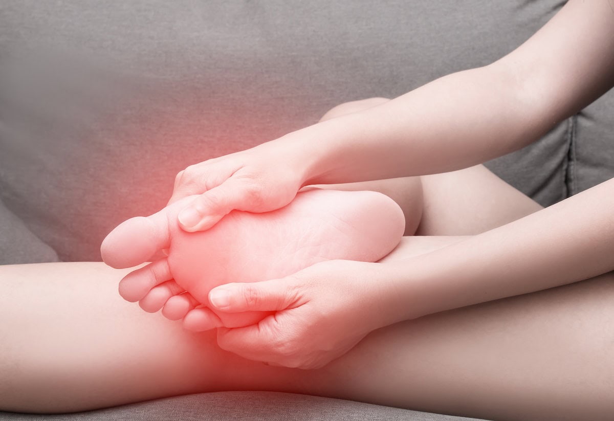
A strained muscle in the leg can happen suddenly during physical activity or build up over time with repetitive use. Whether it’s a mild strain from jogging or a more serious injury while playing sports, knowing how to recognize, treat, and prevent muscle strains is critical for maintaining mobility and avoiding long-term issues. In this blog, we’ll discuss the risk of muscle strains, risk factors, how to reduce swelling, and effective treatments to promote healing and prevent future injuries.

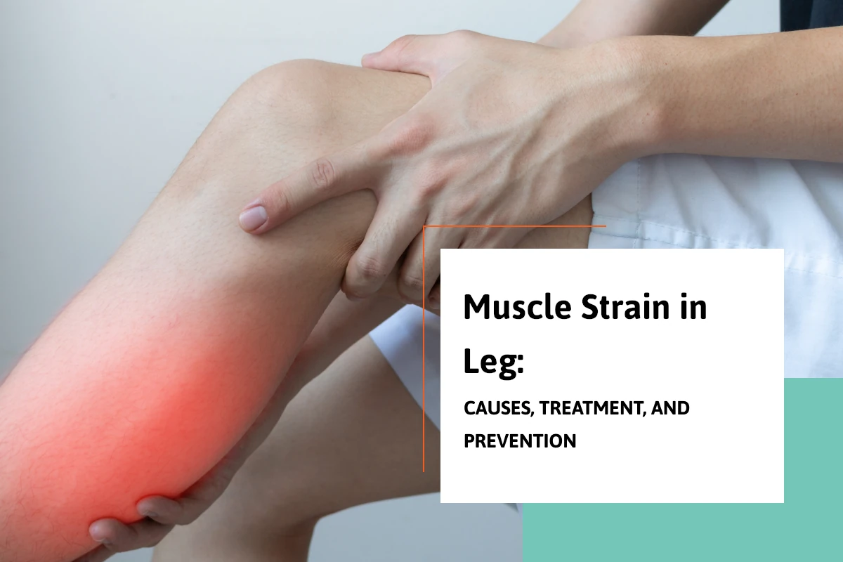
What Happens When a Muscle Strain Occurs in Your Leg?

A muscle strain occurs when the muscle fibers are stretched beyond their limit or even torn. You might feel a pop or sudden sharp pain when the injury happens. This is often accompanied by immediate weakness in the injured area, making it difficult to continue activity. Sometimes, the strain is mild and causes minor discomfort, while other times it can be a complete muscle tear, known as a grade three strain.

Recognizing the Severity of a Muscle Strain In The Leg

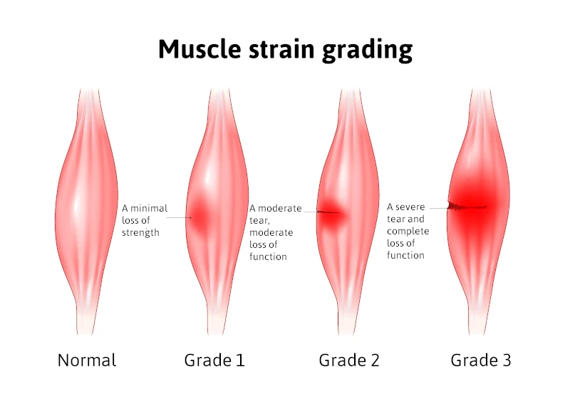
Strains are typically classified into three grades:

  • Mild strain (Grade I): Minor damage to muscle fibers with little loss of strength or motion.
  • Moderate strain (Grade II): More significant injury with noticeable weakness, swelling, and restricted movement.
  • Severe strain (Grade III): Complete rupture of the muscle, often causing intense pain and the inability to use the affected leg.

In some cases, ongoing issues can lead to a chronic muscle strain, where the muscle has not fully healed and continues to cause discomfort during physical activities.

Grades of Muscle Strain In Leg Infographic

Risk Factors for Muscle Strains

Certain factors increase your risk of muscle strains, including:

  • Inadequate warm-up before exercise
  • Poor flexibility
  • Muscle fatigue
  • Previous muscle injuries
  • Age-related changes to muscle elasticity
  • Participation in high-intensity sports

Understanding these risk factors can help you take steps to protect yourself during activities.

How to Prevent a Muscle Strain in the Leg

Fortunately, you can take proactive steps to prevent muscle strains. Some of the best strategies include:

  • Proper warm-up and stretching before exercise
  • Strengthening exercises to support vulnerable muscles
  • Staying hydrated and maintaining electrolyte balance
  • Cross-training to avoid overuse of specific muscle groups
  • Gradually increasing the intensity of your workouts

Consistency in these practices can significantly lower the chance that a pulled muscle will sideline you.

Immediate Treatment: Rest, Ice, Compression, and Elevation

When a strain occurs, early treatment is crucial for minimizing damage and speeding recovery. Follow the Rest, Ice, Compression, and Elevation (RICE) method:

  • Rest the injured area immediately to prevent further muscle tearing.
  • Apply ice wrapped in a cloth for about twenty minutes every two to three hours to reduce swelling.
  • Use an elastic bandage for compression to limit inflammation.
  • Elevate the leg above heart level whenever possible to decrease swelling.

These steps are essential within the first 48 hours after the injury.

Medications That Can Help

To manage pain and inflammation, doctors often recommend non-steroidal anti-inflammatory drugs such as ibuprofen. These medications work to reduce both pain and swelling. However, it’s important to use anti-inflammatory medications as directed and not rely on them as a substitute for proper rest and rehabilitation.

Rehabilitation: Regaining Strength and Flexibility

After the initial swelling and pain subside, it’s important to begin gentle rehabilitation exercises. Restoring range of motion without causing additional injury is the goal. Activities might start with light stretching, followed by progressive strengthening exercises. Physical therapy is often beneficial, particularly for moderate to severe strains.

Ignoring a muscle strain or returning to activity too soon can prolong recovery and increase the risk of chronic muscle strain. Taking the time to fully heal ensures the best long-term outcomes.

When to See a Doctor for a Muscle Strain in Leg

Most mild to moderate strains heal well with home care, but there are times when professional medical attention is necessary. Seek help if you experience:

  • Severe pain and swelling
  • Inability to walk or bear weight
  • Numbness or tingling
  • Noticeable muscle deformity

These symptoms could indicate a grade three strain or another serious injury that might require more advanced interventions, such as imaging studies or even surgery.

Long-Term Outlook

With prompt and proper care, most people fully recover from a leg muscle strain and return to their normal activities. Severe cases may take several months, while mild strains often heal within a few weeks. Adhering to treatment recommendations and rehabilitation plans is essential for preventing a recurrence. Once healed, it’s wise to incorporate injury prevention strategies into your daily routine. This not only minimizes the risk of muscle strains in the future but also helps enhance overall performance during physical activity.

Conclusion

A strained muscle leg injury can be frustrating, but understanding the injury process and knowing how to respond makes a huge difference in recovery. Remember that the risk of strains is higher if you have certain risk factors, but proper preparation and caution can prevent muscle strains. Treat early with rest, ice, compression, and elevation, and support healing with anti-inflammatory medications like ibuprofen when appropriate. Always listen to your body — if you feel a pop or experience intense pain, take it seriously. Healing takes time, but with patience, you can fully recover and return to your favorite activities stronger than ever.

Schedule Your Comprehensive Foot Evaluation Today

Call us today at our Fort Collins location (970) 484-4620, Broomfield location (720) 259-5053 or use our online scheduling system to book your appointment.

With convenient clinical foot locations in Broomfield and Fort Collins, Colorado.

The Hidden Truth About Calcaneal Spur Surgery: What Your Doctor May Not Tell You

calcaneal spur surgery

A calcaneal spur, often referred to as a heel spur, is a bony growth that develops on the heel bone (calcaneus). This condition is commonly associated with plantar fasciitis, which involves inflammation of the plantar fascia ligament, leading to significant heel pain. While many individuals manage heel pain through conservative treatments, some may require calcaneal spur surgery to alleviate persistent discomfort and restore mobility. This blog aims to provide a better understanding of why the removal of the heel spur is rarely needed and can be considered a fallacy. I will contrast the old-school ideas with the more recent concepts based on science.

What is a Calcaneal Spur?

A calcaneal bone spur is a bony growth that forms on the underside of the heel bone, typically at the site where the plantar fascia attaches to the calcaneus. Most believe that the point of the fascia from the heel bone at this location may stimulate new bone to form, thus causing the spur formation. In the 1930s, Dr. Spitz was the first doctor to suggest that the actual heel spur is not causing the pain, but rather the fascia that is pulling from the calcaneus.

This condition can arise due to repetitive stress and strain on the foot, often resulting from activities such as running, walking, or standing for long periods. The bone spur development can occur alongside plantar fasciitis, leading to significant heel pain and discomfort. Symptoms may include sharp pain in the heel, especially in the morning or after prolonged periods of sitting. While many individuals experience relief through non-surgical interventions, some may require surgery when conservative treatments fail to provide adequate relief.

Causes of Calcaneal Spurs

calcaneal spur surgery causes infographic

Calcaneal spurs commonly develop due to a combination of factors, including:

  • Repetitive Stress: Activities that involve excessive strain on the feet, such as running, dancing, or standing for extended periods, can lead to the formation of a heel spur.
  • Plantar Fasciitis vs Plantar Fasciosis: the old school concept is that the fascia is inflamed. This has been disproven by one study performed by Dr. Harvey Lamont. When he performed surgery to release the plantar fascia from the heel bone, specimens were sent to a lab. Out of 50 specimens, 49 showed fascial degeneration while only one showed inflammation. This supports that it is not an inflammatory condition.
  • Chronic pulling of the plantar fascia can contribute to the development of a calcaneal spur, as the body attempts to stabilize the area by forming new bone.
  • Foot Structure: Abnormalities in foot structure, such as flat feet or high arches, can increase the risk of developing heel spurs.
  • Age and Weight: Excess body weight can contribute to higher stress levels on the feet.

Symptoms of Calcaneal Spurs

Individuals with a calcaneal spur often experience a range of symptoms, including:

  • Heel Pain: The most common symptom is a sharp, stabbing pain in the heel, especially during the first steps in the morning or after prolonged sitting.
  • Swelling and Inflammation: The area around the heel may become swollen and tender to the touch due to inflammation.
  • Pain with Activity: Activities like walking, running, or standing for long periods can exacerbate heel pain.
  • Difficulty in Movement: In severe cases, individuals may find it challenging to walk or engage in physical activities due to discomfort. Understanding these symptoms is crucial for individuals to seek appropriate medical evaluation and treatment.

Diagnosis of Calcaneal Spurs

Diagnosing a calcaneal spur typically involves a thorough evaluation by a healthcare professional. The diagnostic process may include:

  • Medical History: The doctor will inquire about the patient’s symptoms, pain levels, and any previous foot injuries or conditions.
  • Physical Examination: A physical examination of the foot will help assess tenderness, swelling, and range of motion.
  • Imaging Tests: X-rays are often used to confirm the presence of a calcaneal spur and assess its size and location. In some cases, MRI or ultrasound may be utilized to evaluate soft tissue involvement, such as the plantar fascia and the Achilles tendon. An accurate diagnosis is essential for determining the most appropriate treatment options, whether conservative or surgical.

Non-Surgical Treatment Options

Before considering surgery, healthcare providers typically recommend conservative treatment options for calcaneal spurs, including:

  • Rest and Activity Modification: Reducing high-impact activities and allowing the foot to heal can alleviate symptoms.
  • Physical Therapy: A physical therapist can design a personalized program that includes stretching and strengthening exercises for the foot and ankle, improving flexibility and reducing pain.
  • Orthotic Devices: Custom orthotics or shoe inserts can provide additional support and cushioning to the foot, helping to redistribute pressure away from the heel.
  • Pain Medication: Over-the-counter medications, such as ibuprofen or acetaminophen, can help manage pain and inflammation.
  • Cortisone injection vs Regenerative medicine: Corticosteroid injections may be helpful but rarely provide permanent relief and this is because the fascia is not inflamed, but rather degenerating based upon studies. Therefore, the option of regenerative medicine provides a very success rate to reverse the symptoms on a long-term basis as this treatment helps repair the fascia that is degenerating.

When is Surgery Necessary?

Surgery for calcaneal spurs is generally considered when conservative treatments fail to alleviate pain and improve mobility after several months. Indications for surgery may include:

  • Persistent Pain: Severe, ongoing heel pain that significantly affects daily activities and quality of life.
  • Diagnosis Confirmation: Confirmation of a calcaneal spur through imaging tests and persistent symptoms despite conservative management.
  • Functional Limitations: Difficulty participating in work, recreational activities, or exercise due to heel pain. Surgical intervention aims to remove the spur and address any accompanying issues, such as plantar fasciitis, to restore function and reduce pain.
  • Regenerative Medicine: The option of regenerative medicine has greatly reduced the need for surgery. (see testimonial videos)

The Surgical Procedures for a Calcaneal Spur

Contrasting heel spur surgery to endoscopic plantar surgery. Calcaneal heel spur surgery usually involves incision on the side of the heel and the spur is removed along with the attachment of the fascia to the heel bone at the spur location. This is a procedure that surgeons have not performed since 1990. Because the heel spur is not thought to be the cause of your pain, using a small incision to place a scope across the bottom of the heel allows the surgeon to cut a portion of the fascia so that it may stretch and elongate and reduce its pull on the heel bone. This technique has been adopted by majority of the Podiatric Foot and Ankle Surgeons, and because of its high success rates, it supports that fact that the heel spur is not the cause of your pain.

Recovery from heel spur surgery

This type of surgery usually involves a larger incision and more trauma and longer recovery with immobilization in a cast, and sometimes non-weight bearing.

Recovery from endoscopic heel surgery

This type of surgery uses a small incision, and you can bear weight immediately and may return to wearing shoes in 3-4 weeks.

Watch Patient Testimonial Videos


Post-surgery, patients are typically advised to avoid weight-bearing activities for a specified period to allow for proper healing. Physical therapy may be recommended to facilitate full recovery and regain strength and range of motion.

Recovery and Rehabilitation for Calcaneal Spur Surgery

The recovery time for calcaneal spur surgery varies among individuals, with most patients experiencing significant improvement within a few weeks. A typical recovery process includes:

  • Pain Management: Pain may be managed with prescribed medications and the application of ice to reduce swelling.
  • Physical Therapy: Engaging in physical therapy is crucial for restoring strength, flexibility, and balance. Therapists may focus on exercises to improve range of motion and stability, particularly around the Achilles tendon and plantar fascia.
  • Gradual Return to Activity: Patients are typically encouraged to gradually return to normal activities as healing progresses, following their surgeon’s recommendations.
  • Long-Term Management: Maintaining a healthy weight, wearing appropriate footwear, and incorporating regular stretching and strengthening exercises can help prevent future heel issues.

Conclusion

Calcaneal spur surgery can provide significant relief for individuals suffering from persistent heel pain due to calcaneal spurs. While many patients find success with conservative treatments, surgery remains an effective option for those who do not respond to traditional methods. Understanding the causes, symptoms, and treatment options for calcaneal spurs is essential for making informed decisions about care. If you suspect you have a calcaneal spur or are experiencing persistent heel pain, consult with a healthcare professional to explore the best treatment options for your needs.

Bunion Surgery Recovery: A Complete Week-by-Week Breakdown

Bunion Surgery week by week

Bunion surgery, or a bunionectomy, can be a life-changing procedure for individuals suffering from painful, misaligned toes. While the surgery itself is relatively common, many patients are unsure about what to expect during recovery. Healing from bunion surgery takes time, and understanding the recovery process week by week can help you prepare both physically and mentally. Here’s what you can expect after bunion correction surgery.

Bunion Surgery Recovery Basics

Before you read the week-by-week recovery that you would expect from bunion surgery, some basic principle bunions need to be understood. With very few exceptions when surgery is performed, the first metatarsal bone needs to be shifted towards the second metatarsal bone to correct the increased angle between the two bones.

This increased angle is what causes the bunion protrusion. This requires that the bone heals in a more correct position. The surgical technique to shift the bone can take place at the level of the metatarsal head which is close to the big toe joint or back at the first metatarsal base which is towards the mid-portion of your foot. Approximately 90% of surgeries we perform are in the area of the metatarsal head and most patients can return to regular shoes between 3 and 4 weeks after surgery. For more severe bunions, they may have to be off weight bearing for 5-6 weeks. This is important to understand because when the bunion starts to hurt, it may be advantageous to get it corrected sooner than later.

The majority of these surgeries take place in an out-patient setting and no general anesthesia is needed. The surgery can be done in a minimally invasive approach and may take approximately an hour and local anesthetic, and IV sedation is given during the surgery. The recurrence rate after the surgery is extremely low, and the patients are able to return to their normal daily activities after weeks post-surgery.

(Watch Bunion Surgery Patient Testimonial Videos)


Week 1: Immediate Post-Surgery Care

The first week is all about rest and protecting your affected foot. You’ll likely experience swelling, pain, and discomfort, which is managed with prescribed pain medication and keeping your foot elevated.

Key Instructions:

  • Keep your foot elevated as much as possible (above heart level) to reduce swelling.
  • Use ice packs periodically to manage pain and inflammation.
  • Avoid putting weight on the affected foot; use crutches or a walker as advised.
  • Keep the surgical dressing dry and intact to prevent infection.
  • Follow up with your surgeon for post-operative instructions.
  • Be aware of deep vein thrombosis risks and watch for unusual swelling or pain.

Week 2: Early Healing and Mobility Adjustments

By the second week after surgery, some pain and swelling may persist, but you should notice gradual improvement. Your stitches may be removed during this period, depending on the types of bunion surgery performed.

Key Instructions:

  • Continue keeping your foot elevated when sitting or lying down.
  • Start moving your big toe slightly if recommended by your doctor to maintain range of motion.
  • Some patients may transition to wearing surgical shoes instead of using crutches.
  • Watch for signs of infection, such as excessive redness, warmth, or drainage.

Week 3-4: Reduced Swelling and Increased Activity

At this stage, you may start feeling more mobile, but your foot and ankle are still healing

Key Instructions:

  • Swelling may still be present, but it should be decreasing.
  • Depending on the procedure, you may begin some weight bearing on the affected foot.
  • You might start gentle range-of-motion exercises to prevent stiffness.
  • Avoid prolonged walking or standing.
  • Keep wearing your surgical shoes as directed.

Week 5-6: Transitioning to Normal Activities

This is when many patients begin to feel significantly better and may even start wearing regular shoes.

Key Instructions:

  • You may transition to a more supportive shoe with a wide toe box.
  • Swelling is still common, especially by the end of the day.
  • You may be cleared for physical therapy to improve strength and mobility.
  • Avoid high-impact activities like running or jumping.

Week 7-8: Resuming Light Exercise and Daily Routines

At this point, many patients find they can walk more comfortably, though swelling may persist.

Key Instructions:

  • Walking distances can gradually increase.
  • Light stretching and strengthening exercises may be introduced.
  • Swelling may still occur, especially after long periods of standing.
  • You might be able to return to work if it doesn’t require extensive foot use.

Week 9-12: Near Full Recovery

By the end of three months after surgery, most patients can return to normal activities with minimal discomfort.

Key Instructions:

  • You should be able to walk normally in comfortable footwear.
  • Continue exercises to maintain mobility and strength.
  • Some residual swelling may persist but should be much less frequent.
  • High-impact activities may be gradually reintroduced with your doctor’s approval.

Month 4-6: Long-Term Healing

Though you may feel fully healed, complete recovery from bunion surgery can take up to a year.

Key Instructions:

  • Continue to wear supportive footwear to prevent recurrence.
  • Regular stretching and strengthening exercises help maintain flexibility.
  • Swelling should be minimal or nonexistent.
  • High-impact sports and activities can typically resume.

Final Thoughts

It is important to understand that most patients we see for bunions come to us because they are painful, and by that time, the conservative treatment options are less likely to be recommended. If the bunion deformity is dealt with earlier before there is pain, orthotic devices (custom-made arch supports) can help with the discomfort and have some effect on slowing the progression of the condition. Unfortunately, most people allow their pain to be their guide and don’t come in earlier where orthotic treatments could still be useful. Orthotics are also useful after the surgery especially in younger people to alter the biomechanics of the foot that created the bunion to begin with.

Recovery from bunion surgery varies from person to person, but patience is key. Following your doctor’s instructions, keeping up with physical therapy, and taking care of your feet will ensure the best possible outcome. If you experience unusual pain, swelling, or difficulty moving your foot, consult your surgeon promptly.

With the right approach, you’ll be back on your feet—pain-free—before you know it!


Stop Putting Up with Your Leg Pain and Treat the Cause!

causes of leg pain

Leg pain is a common complaint that can include a multitude of causes. The purpose of this blog is to give you the more common causes so that you may better understand your leg pain. Generally speaking, leg pain can be caused by injury, overuse, and also from general health conditions. It can also be related to circulatory problems and nerve conditions. This blog may serve as a general guide to know what type of specialist you should see that can treat leg pain based upon which of the following symptoms you may have.

Circulatory conditions to be aware of for causes of leg pain

Circulatory conditions that may cause pain in the legs include varicose veins, thrombophlebitis (blood clot), and peripheral arterial disease.

  • Varicose Veins – This is a common condition caused by veins that do not function properly. Arterial blood flows down the extremity to the foot and has to travel back to the heart through the veins. There are one-way valves in the veins that oftentimes work improperly, and their function is to not allow the backflow of venous blood back toward the foot. When these veins are dysfunctional this can lead to a buildup of venous blood in the veins causing them to enlarge. These are called varicose veins and they can be unsightly and painful.
  • Thrombophlebitis (Blood clot) – A blood clot may occur when the blood in the vein traveling back up to the heart becomes clotted. This can happen in the superficial veins in the lower leg closer to the skin and generally will cause pain in the area of that vein. As the veins travel back up towards the heart, they travel deeper into the leg, especially when they approach the knee joint area. If the deeper veins become blocked with a blood clot this is called deep vein thrombosis (DVT). This will cause more generalized pain in a larger area and possibly the entire leg and it is critical you seek medical attention. If the blood clot breaks off and travels to the heart and lungs, this situation can be fatal. Situations that can lead to this are associated with long periods of sitting such as in an airplane or car for long periods of time, and can be associated with trauma in the lower extremity or after surgery in the lower extremity where there is immobilization and reduced activity.
  • Peripheral Arterial Disease (PAD) – Peripheral Arterial Disease simply means the blood flow from the heart to the foot can be blocked by the narrowing of arteries. This can also be called arteriosclerosis. When the blockages become more severe, pain can occur in the legs with activity, and this is called intermittent claudication. A typical complaint would be that you get muscle cramps and pain in the calf area after walking one or two blocks. You discontinue walking, the pain goes away but when you start walking again the pain returns. This can be an indication of poor circulation. This is more common in diabetics and also the narrowing of the arteries in diabetics tends to happen in the lower leg and foot rather than the larger arteries in the knee area and above. Lack of arterial blood flow to the foot makes you susceptible to infection and even amputation if an injury or infection occurs in the lower leg.

common-causes-of-leg-pain infographic

Nerve symptoms

Nerve symptoms in the legs are very common and the majority of these symptoms are associated with three major diagnoses. The symptoms are the lower back, neuropathy, and restless legs.

  • Lower Back – Many patients are diagnosed with nerve symptoms that are occurring in the leg below the knee and into the foot and it is assumed that the region of the lower back called the lumbar area is causing these symptoms. The area of the lower back called the lumbar area is the area where the sciatic nerve which is the primary nerve that supplies the legs and feet originates. Often patients are diagnosed with radicular pain that radiates from the leg, thigh, and down into the foot. These symptoms can include burning, tingling, and sharp radiations of pain. Treatments for this are directed at treatment of the lower back including chiropractic treatments, physical therapy treatments, injections, and sometimes surgery. The symptoms in the legs would then correspond to lower back pain for most people.
  • Neuropathy – Nerve pain from neuropathy can include burning, tingling, numbness, and sometimes weakness predominantly from the knee down. Oftentimes it can be limited to the top and bottom of the foot and with very little extension to the leg but it can also include the leg. Because conventional medicine has so many healthcare professionals evaluating and treating the back, oftentimes a patient will be told that the neuropathy symptoms are related to their back. As a peripheral nerve surgeon, I see this quite frequently. The patient is led to believe the neuropathy is caused by their lower back when in fact the symptoms are attributed to compression of tight nerve tunnels in the lower extremity. There are five of these nerve tunnels much like the carpal tunnel in the upper extremity. Patients are often told when they have neuropathy whether it is nondiabetic or diabetic it is irreversible.This is not true.As a peripheral nerve surgeon who performs surgery on patients with diabetic neuropathy and nondiabetic neuropathy, we find that the majority of these patients have compression of various nerve tunnels in the lower extremities. There is hope for the reversal of neuropathy! These patients may also suffer from a condition called drop foot which means they have difficulty bringing their foot upward, moving their foot sideways from the opposite foot. They may also have severe balance and gait disturbances because of the numbness combined with their weakness.Conventional treatment is primarily directed at pharmaceutical drugs that manage the symptoms but do not address the cause. (please consider viewing testimonial videos of the reversal of neuropathy and two peer-reviewed papers showing intra-operative nerve testing and objective evidence of nerve function improvement in the operating room)
  • Restless legs syndrome – Restless legs symptoms can be mild or very devastating to patients as it causes patients to lose their ability to sleep, making it difficult to go to sleep, or because of the symptoms they are consistently waking them up. The symptoms include anxiousness in the legs that seems to be present more at night when lying down or during the day when sitting. Symptoms are usually relieved by standing or walking. Other symptoms may include creepy crawling sensations, pulling sensations, cramping, and jerking. Conventional treatments may include the use of iron supplements as low iron levels are thought to be associated with restless legs or taking drugs that increase dopamine levels as the neurotransmitter dopamine is speculated to have lower concentrations in the brain in those with restless legs.As a peripheral nerve surgeon, I have been having great success with nerve decompression surgeries to open up tight nerve tunnels in the lower extremity that are believed to be causing restless legs symptoms. I have published a book called “A Perfect Night’s Sleep that explains my background and the concept that restless legs may be more of a mechanical problem caused by tight nerve tunnels. I also have one peer-reviewed published paper documenting my findings.


Soft Tissue Problems that cause leg pain

This may include the following:

  • Achilles tendinitis or Achilles tendon tear
  • Baker’s cyst – a cyst that occurs in the upper part of the leg just below the knee.
  • Muscle tear – this can happen from an injury, especially to the gastrocnemius muscle.
  • Muscle strains – this can be due to overuse.
  • Cellulitis – this will usually be associated with redness and heat, and it is a soft tissue infection.
  • Shin splints – this can occur from overuse of the muscles that originate in the leg bones that extend down to the foot and are used to stabilize the ankle.
  • Tendonitis – this can be from the Achilles tendon or other tendons around the lower leg and ankle level that stabilize the foot.
  • Growing pains – this can occur in children as they are growing. The child may complain of leg cramps. The idea is that their bones grow faster than their soft tissue structures.

Bone

  • Fractures – The two main bones of the lower extremity – the femur or tibia – may have a fracture. If a significant fracture is present, the pain is severe. If stress fractures are present, pain may be moderate and pain may be localized to where the stress fracture is located.
  • Bone Tumor – Malignant bone tumors are rare, but they can cause significant pain.

Miscellaneous

  • Electrolyte Imbalance – this can be caused by a lack of sodium, potassium, or calcium.
  • Charlie Horse – this often occurs at night and may be associated with electrolyte imbalance or overuse of the lower leg muscle. Gently stretching can help alleviate the Charlie horse.
  • Muscle spasms – this can occur from overuse of the muscles of the lower leg.

Finally, it is important that if you have had leg pain for a very long time, you should see a professional who can evaluate the causes, and treat your leg pain. This blog should help you understand which type of doctor to see based on your symptoms. I should stress that if you have symptoms that seem to be restless legs or neuropathy, you should consider seeing a peripheral nerve surgeon as they may be more likely to get to the root cause of your problem and not just manage your symptoms with medication.

Call us today at our Fort Collins location (970) 484-4620, Broomfield location (720) 259-5053 or use our online scheduling system to book your appointment.

Heel Pad Syndrome: Is it Fallacy or Is it True?

heel pad syndrome

Heel pain is one of the most common complaints that foot specialists encounter. Many people assume that pain in the bottom of the heel is caused by plantar fasciitis, which is true for most patients. However, another less common issue, known as heel pad syndrome, can also cause heel pain. In this blog, we will discuss the causes and treatment of heel fat pad syndrome. Additionally, you may have received a misdiagnosis, and we will explore a little-known condition that could explain why your treatments have not been successful.

The Basics: Heel Anatomy

The foot is uniquely designed to absorb shock. As we walk or run, the foot strikes the ground with 3-4 times our body weight. To compensate for this, the heel, the softest bone in our body, dissipates that shock. Beneath the skin, a significant amount of fatty tissue is distributed throughout the bottom of the foot, providing additional cushioning.

Within the fat are tight connective tissues that act as a net to support the fat pad’s structure. On the bottom of the heel, x-rays may reveal a heel spur. Using ultrasound technology, a podiatrist can also detect abnormalities in the plantar fascia.

The plantar fascia originates in the ball of the foot, extends through the arch, and inserts at the bottom of the heel. With every step, the arch flattens, causing the fascia (a tight connective tissue) to pull on the heel.

heel pad normal foot

Symptoms of Heel Pad Syndrome

When one is suffering from heel pad syndrome, the pain will be directly underneath the heel bone. Pain may be present first thing when you get up in the morning or when you walk throughout the day. The pain will not extend to the arch but is centered directly underneath the heel.

Causes of Heel Pad Syndrome

  • Heredity – Some people may inherit a thinner fat pad from their parents.
  • Cortisone Injections – Cortisone injections, which doctors may use to treat plantar fasciitis, can lead to heel pad syndrome and contribute to fat pad atrophy.
  • Hard Surfaces – Walking or running barefoot, as well as engaging in activities on hard surfaces like concrete or wooden floors, can cause the fat pad to thin.
  • Biomechanics – Certain foot structures are more likely to cause fat pad atrophy.
  • Excess Body Weight – People who carry more body weight may increase their risk of developing heel pain. The added weight stresses and displaces the fat pad.
  • Age – As we age, the fat pad naturally thins as part of the aging process.
  • Metabolic Problems – Medical conditions like type 2 diabetes, lupus, and rheumatoid arthritis can increase the risk of developing heel pain.

Diagnosis of Heel Pad Syndrome:

  • Symptoms – The doctor asks you about your symptoms, where they are located, and when they occur.
  • Examination – The doctor examines your foot to determine where the pain is located.
  • Testing – The doctor may take X-rays to rule out any bony involvement.
  • Diagnostic Ultrasound – The doctor uses ultrasound to measure the thickness of the fat pad and evaluate the plantar fascial tissue in the affected area.
  • MRI Imaging – The doctor may order an MRI to assess the fascia and fat pad, and more importantly, to rule out a stress fracture in the heel.

Treatments may include the following:

  • Shoe Gear – Wearing shoes with extra cushioning can help provide relief.
  • Heel Cups – It will help center the fat directly beneath the heel, preventing displacement from the middle when you bear weight.
  • Medication – Taking anti-inflammatory medications can help reduce pain and inflammation.
  • Icing – Applying ice can alleviate symptoms in mild cases of heel pad syndrome.
  • Shoe Inserts – Using over-the-counter arch supports or custom-made orthotics can offer additional support and comfort.

Life style Changes:

  • Avoid High-Impact Activities – Avoid high-impact activities like basketball that cause heel pain. Jumping and landing on your feet wear down the fat pad. Instead, opt for low-impact activities that involve softer surfaces like swimming.
  • Maintain a Healthy Weight – Keeping a healthy weight reduces the impact and force on your feet, allowing for better distribution and control.

Why the diagnosis of Heel Pad Syndrome should throw up a red flag in your mind?

  • Plantar Fasciitis – When it comes to pain in the bottom of the heel, the most common diagnosis is plantar fasciitis. To accurately diagnose this foot problem, patients with heel pain should undergo a diagnostic ultrasound. Using an ultrasound, a podiatrist can evaluate both the appearance and thickness of the fascia to confirm the diagnosis. Without this test, a definitive diagnosis cannot be made.
  • Heel Spur – Heel spur pain is a common misconception. In the past, people believed that if a heel spur appeared on an x-ray, it was the source of pain. However, with the introduction of endoscopic surgery in the late 90s, this belief has become outdated. This advanced technique, which has proven highly successful in eliminating heel pain, changed the understanding of heel spur pain.

    During this procedure, surgeons make small incisions on both sides of the heel, allowing them to visualize and cut a portion of the fascia to relieve tension. Interestingly, the success of this technique does not involve removing heel spurs, even though many patients undergoing the procedure have heel spurs. As a result, the idea that heel spurs cause pain has been invalidated. In fact, as early as the 1930s, medical literature stated that heel spurs develop due to the fascia pulling away from the heel, but the spur itself does not cause pain.

    Your Fat Pad Syndrome Pain may truly be Nerve Pain. Are you misdiagnosed? Your Fat Pad Syndrome may be Nerve Pain.

The Medial Calcaneal Nerve Branch

The medial calcaneal nerve branch passes beneath the heel bone, located precisely where the pain occurs. This nerve originates from the posterior tibial nerve, which travels along the inside of your ankle. The posterior tibial nerve has branches that supply sensation to the bottom of the foot, much like the carpal tunnel area in the hand. Specifically, two branches in the tarsal tunnel extend to the arch and ball of the foot, providing sensation to these areas.

Meanwhile, the third branch, the medial calcaneal nerve, travels down the inside of your heel and supplies sensation to the bottom of the heel bone. On its way to the heel, it passes through a small tunnel on the inner side of the heel bone. This tunnel, composed of tightly connected tissue, can entrap or compress the nerve. Additionally, when the plantar fascia thickens in patients with plantar fasciitis, this nerve often becomes irritated. As a result, a patient may experience both plantar fasciitis and nerve pain simultaneously.

Diagnosis of Medial Calcaneal Nerve Branch Pain

      • Clinically – The doctor evaluates the heel by checking for discomfort, which usually occurs on the inner side of the heel bone.
      • Diagnostic Injections – The doctor may administer an injection at the entrapment site of the nerve. The location is on the inside of the heel and then it follows beneath the heel bone. When patients respond favorably with a significant reduction in pain, this confirms the diagnosis.

Treatment

      • Cortisone Injections – Doctors can use cortisone injections to help confirm the diagnosis, and a series of two or three injections can eliminate the pain.
      • Radio Frequency Nerve Ablation Technique – This technique effectively blocks the pain signals in the nerve.
      • Surgery – As a last resort, surgeons can resect the nerve and bury it into the muscle in the lower leg.

Have you been told you have heel pad syndrome and the attempts by your doctor have failed? Please consider the little-known diagnosis of medical calcaneal nerve branch entrapment. This may truly be what is slowing you down and causing your heel pain. This is why you should contact our certified foot doctors.

Call us today at our Fort Collins location (970) 484-4620, Broomfield location (720) 259-5053 or use our online scheduling system to book your appointment.


Arch Pain in Foot: The Persistent Causes and Treatment your Doctor Missed

arch pain in foot

Pain in the arch of the foot is a very common complaint seen by podiatrists. One of the most common causes is plantar fasciitis. This blog will discuss many other potential causes of arch pain that you may not have heard of. If you are having arch pain and are frustrated with what you have tried or what your doctor has tried and this problem persists, this blog is for you. We will go into more detail as to other causes that may lead to arch pain. It is important to get to the root cause for your arch pain to resolve.

The Basics of Arch Pain in Foot

Let’s first explain what the arch on the bottom of the foot is composed of.

  • Plantar Fascia. Beneath the skin is a strong connective tissue that goes from the ball of your foot to your heel, and it is called your plantar fascia. As we get older, the fascia becomes less elastic but as a young person, it has more elasticity and if we step down on the foot the fascia has the ability to elongate. This flexibility is lost over time resulting in the pulling of the fascia from where it attaches to the heel bone.
  • Nerves. Deeper to the fascia are multiple nerve branches that supply muscles in the arch and also the skin. The nerves originate from your back and on the pathway to get to the bottom of your feet, the nerves pass through two nerve tunnels. One nerve tunnel is called the soleal sling in the upper calf and another nerve tunnel in the inside of the foot, in the ankle region is called the tarsal tunnel.
  • Musculoskeletal. The musculoskeletal aspect of your foot refers to the muscles, the tendons, the bones, and joints. These could also be the cause of arch pain.

Why am I getting arch pain?

There are multiple things that you could have done that lead to pain in the arch. These include the following:

  • Shoes that have been overused and are breaking down. Wearing shoes that do not provide adequate support can cause multiple problems in your feet. This may occur because the shoes do not provide adequate support or have broken down.
  • Overtraining. Maybe you are starting to do more rigorous activities such as running, walking for more days a week, or hiking. It is important to have rest days between the days you are stressing your body or do alternative activities on those days so that you are not overstressing your feet. When you start to feel pain, using ice packs to apply ice and taking anti-inflammatories may be helpful to relieve pain and reduce swelling if you are overtraining. Icing can be done for up to 20 minutes at a time.
  • Foot type. Some feet structurally are more susceptible to arch pain, and this can include flat feet but also a very high arch foot. Both may lead to arch pain.

Specific Causes and Treatments for Arch Pain in Foot

Plantar Fasciitis

Plantar fasciitis may be the most common cause of arch pain, and it is the most common terminology used by the public and healthcare professionals. However, because of this, the diagnosis may be overused and especially if you are not seeing a foot specialist and not getting better, you should start to question the diagnosis, and you may want to consider getting a second opinion.

Where is Plantar Fasciitis located?

With very few exceptions, plantar fasciitis is closely located where it is inserted in the heel. People present with pain in the bottom of the heel when they first get up in the morning and bear weight. As the arch flattens, the plantar fascia is pulled, and it is believed that microscopic tears can be created at the juncture where the fascia attaches to the heel bone. It may also occur in the middle of the foot, but it is much less. This is important to note because if the pain is in the middle of the foot, other problems may be the cause.

Treatment for Plantar Fasciitis

Treatment can involve more supportive shoes, or a podiatrist may recommend custom-made supports called orthotics. Other treatments such as regenerative medicine where human cellular tissue products are used to repair the damaged fascia at its connection of the heel bone can be utilized. Some patients have also found that icing, resting, and using over-the-counter inserts may be helpful. Finally, physical therapy using stretching and strengthening exercises may also be useful. After all conservative measures have failed, endoscopic heel surgery may be performed to release a portion of the fascia so that it can elongate and therefore reduce its pulling from the heel.

Nerve Conditions

Tarsal Tunnel Syndrome

The tarsal tunnel is an area where all the nerves on the bottom of the foot need to pass through to supply the bottom of the foot. It is located on the inner side of the foot below the ankle bone. Much like carpal tunnel syndrome in the hand, if this tunnel is tight, you may get symptoms of burning, tingling, and numbness in a portion of the arch, or in the entire bottom of the foot. Treatments for tarsal tunnel syndrome may include custom-made arch supports, cortisone injections may be used to help diagnose but seldom reverse the symptoms, laser treatments may also be effective. If symptoms are unresolved and severe, surgical decompressing (opening) of the nerve tunnels in that area may be needed to be performed much like doing carpal tunnel in the hands.

Soleal Sling Entrapment

Another nerve condition can create symptoms in the arch from a nerve tunnel located in the upper calf area. The main nerve that supplies the bottom of the foot is called the tibial nerve and it is a branch of the sciatic nerve. It passes through a tunnel that is located where the soleus muscle originates in the upper calf. The soleus muscle which inserts in the Achilles tendon originates in this area and where its origination is a fibrous band that can entrap (compress) the nerve. This band is referred to as the soleal sling. The symptoms from this can create burning, tingling, and numbness of the foot and the arch, and it occurs more with activity and dissipates with less activity. It is also found to cause significant cramping in the arch. Very few health professionals are adequately trained to evaluate and treat this problem, and many are unaware of its existence as a possible cause of arch pain. Treatments for soleal sling entrapment would include laser treatment, electrical stimulation treatments, cortisone injections which may help temporarily, and if symptoms are severe and chronic, surgical decompression (opening) of the tunnels may be needed.

Musculoskeletal

  • Stress Fractures – For some, a stress fracture in the bones of the arch of the foot or the long bones in the foot called the metatarsals can lead to arch pain. Although rare, there are situations where a stress fracture in the arch of the foot can cause cramping of the muscles thus causing pain in the arch. The cramping is present because the body is trying to splint or immobilize the bone because of the fracture. This is nature’s way of attempting to reduce stress on the bone and thus help the bone to heal.
  • Tendons – There are areas in the arch where important tendons attach to the foot. If these tendons become overused there can be mild tears where they connect to the bone. For treatment, it is important to properly diagnose with an ultrasound evaluation or an MRI. In many cases, custom-made arch supports can eliminate the pain and in other situations, regenerative medicine can be used to repair the damage to the tendon or surgery may be necessary.
  • Arthritis – There are multiple joints in the mid arch of your foot that are formed by the tarsal bones and the articulation of the tarsal bones with the metatarsals. As we get older, these joints may become arthritic and the body’s response to arthritis is splinting of the muscles in the arch to immobilize the joints, and this can help reduce the pain. Pain may also be noted on the top of the foot but also on the bottom of the foot in the arch. Treatment for this is more supportive shoes, custom-made arch supports, surgery to fuse the joints, and also regenerative medicine can help reduce joint pain.

Severe Flat Feet

Finally, it must be mentioned that there are situations where a foot may be severely flat and most conservative measures may fail and because of this, a podiatrist may need to consider the option of surgery. With a severe flat foot, there could be severe stress or strains on tendons, nerves, and joints where surgical correction would be the preferred treatment plan.

If you are having arch pain that is getting unresolved, make sure to consider many of the other possibilities besides plantar fasciitis. Plantar fasciitis is a big buzzword in healthcare, and it is important that you are armed with this information so that you can get the best treatment possible for your arch pain if you are not responding to the present treatment plan you are going through. And finally, you may want to consider consulting with a board-certified peripheral nerve surgeon as seeing this type of specialist would be best if you are suspicious of nerve involvement as mentioned in this blog. In summary, please consider visiting The Association of Extremity Nerve Surgeons to find peripheral nerve surgeons in your area.

Book your appointment online or call our Fort Collins and Broomfield, Colorado foot and ankle specialists at the clinic nearest you.

Frequently Asked Questions About Foot Arch Pain

  1. What are the most common causes of foot arch pain?

    Common causes include plantar fasciitis, nerve conditions like tarsal tunnel syndrome, poor footwear, overtraining, and certain foot types like flat feet or high arches.

  2. How do I know if my foot arch pain is due to plantar fasciitis or something else?

    Plantar fasciitis usually causes heel pain when first standing in the morning, while persistent arch pain, burning, or numbness may suggest a nerve issue like tarsal tunnel syndrome.

  3. Can wearing the wrong shoes lead to foot arch pain?

    Yes — worn-out or unsupportive shoes can cause or worsen arch pain by failing to provide the necessary support for your foot’s natural structure.

  4. When should I consider seeing a specialist for foot arch pain?

    If your arch pain persists despite rest, better footwear, and home treatments, or if you’re unsure of the diagnosis, it’s wise to see a podiatrist for a thorough evaluation.

  5. What treatment options are available for chronic foot arch pain?

    Treatments range from orthotics, physical therapy, and regenerative medicine to nerve decompression surgery, depending on the underlying cause.

Call us today at our Fort Collins location (970) 484-4620, Broomfield location (720) 259-5053 or use our online scheduling system to book your appointment.

An introduction to the importance of the lateral cuneiform bone in your foot

lateral cuneiform bone pain anderson podiatry center

The topic of this blog is the lateral cuneiform bone. You probably have never heard of this bone. There are many bones in the human foot, 26 total. Names of foot bones that may be more familiar to you are the metatarsal bones, phalanges, calcaneus (heel bone), and talus (ankle bone). You may have heard of these bones because pain or fractures may occur in their location. The lateral cuneiform bone has a significant role in foot function, however it does its job silently. It does the work it was built to do, and you’ll seldom hear from it as it’s very rare to get injured. It’s part of a group of bones called the tarsal bones that we’ll discuss before diving into the lateral cuneiform bone specifically.

Basic foot anatomy

The foot can be separated into three separate areas. The rearfoot, is comprised of the calcaneus (heel bone), and the talus (ankle bone). The forefoot is composed of the metatarsal bones and phalanges and finally, the area in between is called the tarsal bones.

The tarsal bones. There are five bones between the rearfoot bones and the forefoot bones. Together they form the transverse arch of the foot that supports the arch. Starting towards the ankle there is the navicular bone, on the medial side of the foot, which forms a joint with the talus bone (ankle bone), and the cuboid bone which forms a joint with the calcaneus (heel bone). On the other side of the cuboid bone towards the forefoot are the fourth and fifth metatarsals that articulate with the cuboid bone forming separate joints. The cuboid and fourth and fifth metatarsals form the lateral arch. The navicular bone as we move away from the ankle towards the forefoot has three bones that articulate with the navicular to the first, second, and third metatarsals. These wedge-shaped bones are referred to as the first, second, and third cuneiforms. The medial cuneiform bone is the largest with the second being the smallest. They are also called the medial cuneiform, intermediate cuneifor,m and lateral cuneiforms.

The lateral cuneiform referred to as the external cuneiform,(middle cuneiform) which is the topic of this blog, forms the joint with the navicular bone and also the middle cuneiform medially and lies medial to the cuboid bone. It is also directly connected to the third metatarsal and does have some articulation with the second and fourth cuneiforms sharing articulation with the second and fourth metatarsals. It directly articulates with four bones. Together the four bones we just discussed form the medial arch. This arch compared to the lateral arch has more strength and can withstand more weight.

One of the functions of the lateral cuneiform bone is to serve as an insertion of the tibialis posterior tendon. The posterior tibial tendon is a very important tendon for normal foot function. It’s primary function is the support of the arch and decelerator pronation which means flattening of the foot. With part of the posterior tendons inserted into the foot on the bottom of the third cuneiform, it has an important function to stabilize the arch. Without this tendon, the foot loses much of the support in the arch. It also serves as the insertion of the flexor hallucis brevis muscle which assists in flexing the great toe downward.

Injuries to the lateral cuneiform bone

These are very rare. As I mentioned earlier it silently does its job assisting in supporting your arch. However, there is one type of injury called a Lis franc’s joint injury. Lis Franc’s joint refers to the joints formed by the metatarsal bones and tarsal bones. This injury is caused by a mechanism where the forefoot bones are forced downward in relationship to the tarsal bones. This may happen when landing on the ball of the foot forcing the ankle joint to move downward. The foot is not built well to withstand this type of force and as a result, you can suffer from multiple injuries to the bones including fractures and dislocations. This injury involves the bones and joints that form Lis Franc’s joint. In this situation, there may be an injury to the third cuneiform and its joints.

So don’t get too alarmed if you’re getting pain in your midfoot area as its most likely not from this bone. If needed the specialist may offer x-rays, MRI, or CAT scan to rule it out. However, I hope this blog gave you a better concept of the complexity of the human foot and how well if performs for us on a daily basis. You now understand the importance of the tarsal bones and more specifically the lateral cuneiform.

If you’re experiencing persistent midfoot pain or any other foot-related issues, it’s important to seek professional care. At Anderson Podiatry Center, our specialists are dedicated to diagnosing and treating foot conditions with expertise and compassion. Whether you’re in Broomfield or Fort Collins, our team is here to help you get back on your feet. Don’t hesitate to schedule an appointment today and let us take care of your foot health.

Call us today at our Fort Collins location (970) 484-4620, Broomfield location (720) 259-5053 or use our online scheduling system to book your appointment.

What Is Tibialis Anterior Pain And What Are The Optimum Treatments

tibialis anterior pain

A person may experience tibialis anterior pain and not consider looking into what could be causing the issue. The tibialis anterior tendon is located in the lower leg along the shin bone, tibia. It is the third largest tendon going into the foot with the Achilles tendon being the largest and posterior tibial tendon the second largest. It’s function is to pull the foot up towards the knee. Without it your foot would slap down as you walk as the heel strikes the ground. It also helps to supinate your foot as it helps to support the arch. This tendon is important for normal gait as it is needed for your foot to clear the ground as you walk or run.

Where is anterior tibialis tendonitis pain located?

For the majority of patients, the pain will be above the ankle joint along the shin bone. It may also occur in front of the ankle and down into the foot where it inserts in the foot onto the navicular bone. This bone is halfway between the great toe joint and the ankle joint. It is less common to cause pain in the foot itself.

What causes tibialis anterior pain?

  • Overuse – doing too much too soon can be a cause. Any activity that requires excessive use of the tibialis anterior muscle may be a cause. Because the tibialis anterior muscle is used to decelerate downward motion of the foot at the ankle joint and is used for pulling the foot upwards a the ankle joint motions that may amplify these motions may cause overuse. Example would be hiking on steep terrain as when walking downhill the muscle is used more to decerlate the downward motion of the foot so your foot doesn’t slap the ground. Racquet sports may also be more likely to cause excessive use of the muscle and tendon.
  • Trauma – You may also have a full or partial rupture of the tendon from and ankle injury. This could be caused by a laceration to the tendon or by any force that places excessive downward motion on the foot.
  • Foot types – certain foot types may be more likely to cause tibialis anterior tendonitis. Because the tendon helps to support the arch a flat foot is more likely to place excessive stress on the muscle and tendon.
  • Foot drop – When there is a partial or full rupture of the tibialis anterior tendon a foot drop may develop. Because the primary function of the tendon is to move the foot upwards, if it loses its function the foot will slap against the ground when walking or running. This can lead to ankle sprains as it has a significant effect on balance and stability.

How is tibialis anterior pain diagnosed?

  • Clinical examination– The doctor will find pain and swelling along the shin bone on the front of the leg adjacent to the tibia. Or there will be pain along the tendon in front of the ankle and down into the foot where the tendon attaches to the top of your arch. There may be pain when you pull the foot up in these same areas. If there is a partial or full tear of the tendon it may be difficult to pull the foot upwards.
  • Diagnostic ultrasound– This is a imaging technique that can be used to access the integrity of tendon and muscle.
  • MRI– The doctor may also use MRI which can very accurately evaluate a potential tear and he severity of it.
  • Gait analysis – simply watching you walk may help to rule out foot drop which could be caused by a tibialis anterior tear.

Treatment options for tibialis anterior pain

  • Rest and anti-inflammatories – this can be useful if the pain is not long term and the pain is mild.
  • Immobilization – Using a Cam walker to walk in can also be helpful as it will help rest the tendon and muscle.
  • Modification of activities – Choosing another activity or decreasing the activity that may have contributed to the problem may be recommended.
  • Orthotic devices – Custom made arch supports may help to reduce the stress placed on the tendon as it may be overused in attempting to support the arch of the foot.
  • Physical therapy – different physical therapy modalities may also be helpful.
  • Laser treatment – Use of MLS laser for pain treatment has also been proven successful in our practice for tendonitis issues.
  • Surgery – When a tear or partial tear is present surgery may be recommended to maintain the integrity of the tendon. This type of surgery may require non- weight bearing for minimum of 4 weeks.

Tendonitis versus tendinosis – We now know that what was once thought to be an inflammatory issue , tendonitis may be incorrect and in fact it’s more likely that the tendon is breaking down. There may be small microscopic tears in the tendon. This is often referred to as tibialis anterior tendonopathy. For close to two decades now we have had great success with the use of Human Cellular tissue products.

What are human cellular tissue products?

Sometimes referred as stem cell treatment this can alleviate the need for surgery. When there is severe pain along the tendon, and the MRI or ultrasound reveals tears this treatment can be used. This form of treatment has greatly reduced the need for the surgical repair of tendons in our practice. It is not suggested for a full tendon tear but when the tears are minor or partial it can work extremely well.

How would human cellular tissue products be used?

This is done in a clinic setting. The area where the tendon tear is located is numbed with local anesthesia so it’s a pain free experience. Then with the use of diagnostic ultrasound to locate the area of tendon damage the stem cell product is injected into the area. The patient is allowed to walk immediately and uses a Cam walker to walk in for four weeks.

So consider these options if you have tibialis anterior pain. And one last consideration is to avoid the use of cortisone in the area of the tendon. I do not consider this a good option as it can potentially weaken the tendon and perhaps cause a rupture.

Easily schedule your appointment online or contact our skilled foot doctors at the nearest foot and ankle clinic in Fort Collins or Broomfield, Colorado to find the best podiatrist near you.

Call us today at our Fort Collins location (970) 484-4620, Broomfield location (720) 259-5053 or use our online scheduling system to book your appointment.

Phalanges of the Foot: A Guide to Their Complex Anatomy

phalanges in the foot

There are many bones in the foot making its anatomy one of the more complex in your body. Of the 26 bones in the human foot approximately half of them are phalanges in the foot. Generally, as we start towards your ankle joint and move down the foot towards the toes the number of bones become more multiple in the distal extremity. For example, in your rearfoot there are two large bones the talus (ankle bone) and calcaneus (heel bone) and 14 phalanx bones. The anatomy of the hands and feet are similar but because you walk on your feet one could argue they are more of a structural wonder. We will describe the anatomy of these bones and potential issues that could cause pain in this area of your foot.

Basic anatomy of phalanges in the foot

The phalanges are the bones of the foot that comprise the bone of your toes. They are connected to the larger and longer metatarsal bones that extend from the midfoot down to the ground forming the ball of your foot. When you bear weight on the ball of the foot you’re standing on the metatarsal heads on the plantar surface of these long bones. The phalanges extend beyond the ball of the foot forming the digits. They are comprised of the proximal, middle and distal phalanges.

Proximal phalanges of the feet

These bones form the articulation with the metatarsal bone These two bones form the metatarsal phalangeal joints. These five joints that form the ball of your foot have a lot of upward mobility called dorsiflexion. This allows these joints to bend upwards to allow you to pivot up onto the ball of the foot when you walk or run. The anatomy of these joints is very complex as they require various tendons and ligaments to maintain their stability. This is especially true for the great toe joint. This has beneath it two bones called sesamoid bones that glide beneath the great toe joint as the toe moves up and down, much like your kneecap. The function to improve leverage and therefore strength to the great toe joint as this joint is important to assist in propelling us forward when walking or upward when we jump.

Intermediate phalanges

The next phalangeal bones as we move away from the ball of the foot are the intermediate phalanges. They may also be called the middle phalanges as they lie in the middle of the toes. They are smaller than the proximal phalanges bones. As was just mentioned the great was unique as it has two bones beneath it called the sesamoid bones and there is one more difference with the great toe. The great toe only has two phalanx bones. They are referred to as the proximal and distal phalanx bones.

Distal phalanges

At the very end of each toe are the distal phalanges. These are the smallest of the phalanges and lie beneath the toenails. Rather than having a tubular shape like the proximal and intermediate phalanges they are short and triangular shaped.

The joints of the toes.

We had mentioned earlier that the joint formed by the metatarsal bones and the proximal phalanges is called the metatarsal phalangeal joint.

  • The joint formed by the proximal and intermediate phalanx is called the proximal interphalangeal joint (PIPJ).
  • The joints formed by the intermediate phalanx and the distal phalanx are called the distal interphalangeal joints (DIPJ).
  • Finally, the great toe must be different as it only has two phalanges, so it does not have a proximal and distal interphalangeal joint. Because it has only two phalanges its joint between these two bones is called the interphalangeal joint.
  • Finally, the great toe also has its name, referred to as the Hallux.

To summarize the big toes are different in three ways

  1. It has only two phalanx bones.
  2. It has two sesamoid bones beneath it at the metatarsal phalangeal joint.
  3. It has its own name, Hallux.

Functions of the phalanx bones in the foot

These appendages help with your balance and push your foot off the ground when you walk, run, or jump. The great toe is more important for these purposes than the other four toes. It’s important to note that in a situation of an amputation of a digit. Removal of one of the smaller toes will have little effect on your balance and ability to function normally. However, the absence of the great toe has more impact.

Problems that can arise in the phalanges

Since these bones are the distal extremities of the foot, they can be subject to trauma such as stubbing your toe. When this happens you may fracture a phalanx bone. Contrary to the popular notion that nothing can be done it’s important to have the injured toe evaluated. Why? If a fracture is present a common treatment will be to buddy tape the injured toe to an adjacent toe. This is important during the healing phase to ensure the fracture does not get too displaced. A fracture that heals in a misaligned position can create long-term pain and require surgical intervention. When the great toe is fractured, proper evaluation is needed to ensure proper healing because of its importance for normal ambulation. Again, proper treatment and follow-up is important.

Hammertoes

Hammertoes are deformities of the digits and can create pain from the dorsal surface of the toe rubbing shoe gear of the walking surface. There are three types of hammertoes
Mallet toe. This deformity is at the distal interphalangeal joint. The metatarsal phalangeal joint and proximal interphalangeal joint are in good alignment; however, the distal interphalangeal joint is flexed downward.

Claw toe. This deformity involves all the joints. The metatarsal phalangeal joint is flexed upwards, and the proximal and distal interphalangeal joints are flexed downward. The bases of the middle phalanx may rub shoe gear.

Hammertoe this deformity involves two joints. The metatarsal phalangeal joint is flexed upwards, and the proximal interphalangeal joint is flexed downward.

Treatment for hammertoes is

  • Conservative – wearing appropriate shoes with bigger toe box, padding, etc.
  • Surgery – Foot Surgery is performed to realign the digit to its normal position.

Hopefully, this blog has given you more insight into the phalanx bones, their anatomy, and problems you may encounter in this region of your foot. If you are seeking a ‘podiatrist near me’ in Broomfield or Fort Collins, consider Anderson Podiatry Center. We have our own surgical foot center that has many advantages which you can review here.

Call us today at our Fort Collins location (970) 484-4620, Broomfield location (720) 259-5053 or use our online scheduling system to book your appointment.

What Is Sesamoiditis And What Is The Best Sesamoiditis Treatment?

sesamoiditis treatment anderson podiatry center

The sesamoid bones are small pea shaped bones that sit beneath the big toe joint. Where there is pain under your big toe it may be called sesamoiditis pain. Let’s look at why you may be seeking sesamoiditis treatment to resolve a common foot pain.

Sesamoiditis basics – The sesamoid bones are like the kneecap, patella bone. They sit beneath the big toe joint and the great toe, and both are attached to muscles and tendons that help pull the big toe downward. Much like the kneecap they each glide in a groove under the joint. They function to increase the leverage of the muscles of the foot to pull this toe downward. This motion is very important for normal gait while running or walking. You may have heard of the term turf toe, this is associated with the sesamoid bones but may also involve inflammation to the tendons and muscles that attach to the sesamoids.

What are the symptoms of sesamoiditis?

Sesamoiditis pain tends to be very localized beneath the big toe joint in the ball of the foot. You’ll favor your foot tending to avoid putting pressure on the bottom of the great toe. High heels will make the pain worse. You may have swelling associated with it. The pain tends to be deep and dull, and you may notice it the most when you first step down in the morning. Which is why most people try to pass it off as morning stiffness and not seek sesamoiditis treatment.

What is the cause of sesamoiditis?

  • Foot type – some feet may tend to roll inward referred to as pronation. This would cause more pain to be placed on the ball of the great toe.
  • Trauma, sesamoid fractures. You may have fractured the sesamoid bone. You’ll usually be aware of this as a lot of force would be needed to cause a fracture. Landing with a lot of weight on the great toe or severely jamming your great toe upwards may cause a fracture.
  • Bunions – A bunion deformity causes the great toe joint to be misaligned. Because of this the seamaids may no longer glide centered in the grooves beneath the metatarsal bone but may now be gliding over centered. This may result in erosion of the cartilage on the ridge that separates the two grooves. Both the cartilage on the seamaid bone and the cartilage on the bottom of the metatarsal breakdown causing octopartite to occur
  • Hallux Limitus – This is a deformity of the great toe joint that limits range of motion of the great toe joint. As the joint starts to reduce its motion this may also interfere with the normal gliding of the sesamoid bones and produce pain.
  • Overuse- This may be the most common cause and typically develops gradually. This is especially true in activities that may cause a lot of weight bearing beneath the big toe such as ballet, dancing, basketball and volleyball to name a few.
  • Shoe gear – If you wear high heels excessively it may cause sesamoiditis. Soft soled shoes would be best.

How is sesamoiditis diagnosed?

  • Physical examination – your podiatrist will evaluate your foot to see if you have pain and swelling where the sesamoid bones are located.
  • x-rays- these will be taken to see if the sesamoid bones appear normal.
  • Congenitally abnormal – you may have been born with an enlarged sesamoid or one that is bipartite, has two parts. These abnormal presentations make it more likely to have pain from these sesamoids.
  • Alignment – are the sesamoids positioned where they should be beneath the great toe joint. A bunion deformity will cause them to be misaligned and more likely to hurt.
  • Fracture – the x-rays will also help to rule out a fracture.

How can I avoid sesamoiditis pain?

  • Surgery – Surgery to remove the pain of damaged sesamoid may also be performed but only after conservative measured have failed. When surgery is performed it is usually the medial sesamoid that tends to be more prone to pain. This sesamoid is the one that is located towards the other foot.
  • Reduce or eliminate activities – for some it may be practical to eliminate an activity that causes more stress to this area of the foot. For others better management of how much of the specific activity that is more likely to cause an injury should be considered. You should gradually increase each activity or consider doing it less frequently.
  • Avoid high heels – these will tend to put more weight on the bottom of the great toe joint.

What is the Best Surgical and Non-Surgical Sesamoiditis Treatment?

Non-Surgical Treatments

  • Rest and Activity Modification – Reducing weight-bearing activities to allow the sesamoid bones to heal.
  • Orthotics – Custom orthotic inserts have been shown to have great success by limiting the amount of weight placed on the sesamoid bone to relieve pressure on the sesamoid bones.
  • Padding and Taping – Protects the sesamoid bones and restricts excessive movement. This may help by minimaxing the motion in the joint and thereby reducing the pain.
  • Cam-walker (short leg fracture brace) – By resting and limiting the motion of great toe joint.
  • Anti-inflammatory NSAIDs and Ice Therapy – Helps reduce pain and inflammation in more acute stages where the pain has been short-term and not too severe. Using an Ice pack or wrapped may also help.
  • Physical Therapy – Techniques like ultrasound and laser therapy to promote healing by reducing inflammation. We use the MLS (Multiwave Locked System) to accelerate healing.
  • Corticosteroid Injections – May be used to reduce severe inflammation and pain. When combined with orthotics it has been shown to beeffective.

Surgical Treatments

  • Sesamoidectomy – Removal of the affected sesamoid bone if chronic pain persists despite conservative treatments.
  • Bone Drilling or Grafting – In cases of fractures, drilling may stimulate healing, while severe cases may require bone grafting.
  • Joint Realignment Surgery – If misalignment from a bunion or hallux limitus contributes to pain, realignment surgery may be needed.

Finally, the number one way to reduce the chances of getting sesamoiditis and prevent recurrence

Orthotics you can have your cake and eat it too. The use of custom-made arch supports may be your best option as a sesamoiditis treatment. Especially if you want to do activities that might make you more prone to injury of the sesamoids. These activities may be tennis, pickleball, running, walking, basketball etc. With every step you take you reduce the load on the sesamoid bones. So don’t assume that you’ll always need to limit your activities or stop doing what you’re presently doing.

Call us today at our Fort Collins location (970) 484-4620, Broomfield location (720) 259-5053 or use our online scheduling system to book your appointment.

Frequently Asked Questions (FAQs) About Sesamoiditis

  • What is the best sesamoiditis treatment?
    The best treatment depends on severity. Non-surgical options like orthotics, physical therapy, and NSAIDs work well for most cases. Surgery is reserved for persistent pain or fractures.
  • How long does sesamoiditis take to heal?
    Mild cases can heal within a few weeks with rest and treatment. More severe cases or fractures may take months.
  • Can sesamoiditis go away on its own?
    If caught early and managed with proper footwear and rest, symptoms may subside. However, untreated cases can worsen over time.
  • When is surgery necessary for sesamoiditis?
    Surgery is considered when conservative treatments fail, there is a chronic fracture, or the sesamoid bone is severely damaged.
  • Can I walk with sesamoiditis?
    Walking is possible, but excessive pressure on the forefoot can worsen symptoms. Using orthotics or a walking boot can help reduce strain.
  • Does cortisone help sesamoiditis?
    Cortisone injections can temporarily reduce inflammation and pain, but they are not a long-term cure.
  • What shoes should I wear for sesamoiditis?
    Low-heeled, cushioned shoes with a wide toe box help reduce stress on the sesamoid bones.