Book Now

The Black Spot On Toenail: Are You Worried And What To Do

black spot on toenail

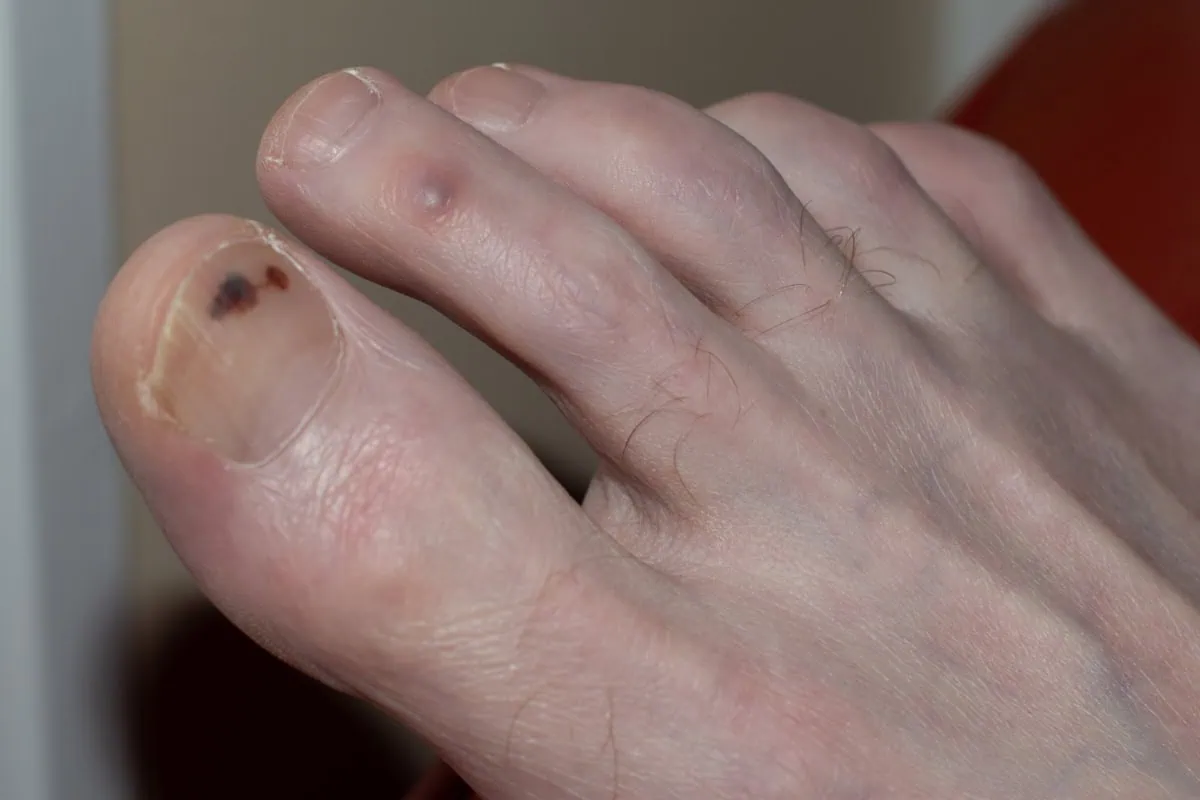
In this blog we will cover what it means if you have black spot on your toenail, whether this is something you should be worried about or something you can ignore, and when to seek medical attention. We will also cover the causes of black spots under the toenail and suggest treatments for black spots that you may want to consider. Black spots and suggest treatment options that you may want to consider.

What can cause a black spot on a toenail?

Fungus

Fungus may be one of the most common causes of blackspot on the toenails. Toenail fungus is more likely to cause yellow discoloration, but there is one form of fungus that’s called tinea nigra that could cause a black spot under the toenails. If the toenail has significant thickening from the toenail fungus, this could lead to increased pressure on the skin beneath the nail especially when wearing shoes that are too tight. The blood vessels in this area break open and lead to bleeding causing a black appearance on the nail bed

Trauma

Trauma to the nailbed may occur from dropping something on your foot may be the cause of a black spot on your toenail. It could also be from minor injuries from when you hike, run, or walk for long distances. The shoe irritation can lead to skin irritation beneath the nail.

Medical Conditions

Certain medical conditions may lead to unhealthy nails such as diabetes mellitus or autoimmune diseases that has a negative effect on the immune system that fight against fungus. Kidney diseases may also lead to the development of a black spot on toenail.

Pseudomonas Infection

This is a type of bacterial infection that can cause green type of discoloration but may also present with brown discoloration associated with it.

Toenail Polish

Generally speaking, toenail polish is not healthy for the nail and could cause damage to the nail which may result in dark patches or discoloration.

Melanoma

Melanoma is a form of skin cancer that may occur underneath the toenail. This is commonly referred to as subungual melanoma. There are many types of skin cancer, but melanoma is the most common type of skin cancer that causes the black appearances on the nail. The area that is blackened will usually run along the length of the nail which is a linear type of presentation and over time tends to expand.

Treatment Options for Black Spots on the Toenail

Surgery

Surgery may be recommended by your podiatrist if the nail has been traumatized, and the cause of the black discoloration is blood beneath it. If a significant amount of nail has been detached from the skin that it is anchored to, it may be advisable to go ahead and remove the entire nail as this will potentially help with the new nail growth so that it will not become deformed. It may also be necessary to relieve the pain associated with this injury. Surgery may also be indicated if melanoma is suspected, and the podiatrist may submit skin tissue sample to a lab for further evaluation for possible melanoma.

If the black toenails are caused by medical conditions, it is important that these medical conditions such as diabetes are managed appropriately as this will improve the general health of the patient and the patient’s immune system. This may also help the body’s ability to fight off fungal or bacterial infections.

Elimination of Toenail Fungus

There are three primary ways that toenail fungal infection can be treated and they are the following:

  • Topicals – There are various topical treatments that can be used that are available over the counter, or you may schedule a visit with your podiatrist and get prescriptions for topicals that may be more effective. Generally, topicals do not work very well and need to be used very in early stages when the toenail is not that severe.
  • Oral medication – Lamisil is a common oral medication that can be used and has more effectiveness than topical medication. However, it is toxic to the liver and many doctors are hesitant using it. It is also important to get your liver health checked before this medication is prescribed.
  • Laser nail treatment – laser nail treatment is the most successful option for eliminating toenail fungus. In this practice, we use a Pinpoint Laser that generates pulses of light into the nail. The light that is pulsing is very hot, but the pulses are occurring so rapidly that most patients have no discomfort and no head is noted. There are no known side effects, and this usually requires a minimum of three treatments sessions.

Melanoma Treatment

If melanoma is suspected, it is important that is it appropriately diagnosed and treated. If left untreated, melanoma can spread to other parts of the body. Treatment of melanoma for most patients involve consultation with a dermatologist who may excise the melanoma and evaluate for possible spread of the tumor and treat appropriately.

One of many ways you can avoid dark spots on your toenails is by keeping your feet clean, dry, and by wearing breathable shoes. Shoes that do not breathe allow for more perspiration in your shoes and this may play a role in increasing the odds of getting toenail fungus which can lead to dark spot on your toenail. It is important to make sure you select your shoe gear appropriately especially when you are very active with activities such as running or skiing. Most importantly, if black spots are found on your toenail and it appears to run the length of the nail and is expanding, it is important to take precautions and make sure you see a foot specialist or a dermatologist to rule out melanoma.

Noticed a black spot on your toenail? Don’t ignore it—early diagnosis is key! Our expert foot specialists in Fort Collins and Broomfield are here to help.

Call us today at our Fort Collins location (970) 484-4620, Broomfield location (720) 259-5053 or use our online scheduling system to book your appointment.


Understanding Tinea Pedis: Symptoms, Causes, and Effective Treatment Options for Athlete’s Foot

Tinea Pedis athlete's foot anderson podiatry-center

Tinea Pedis aka Athlete’s Foot – Anderson Podiatry Center

Athlete’s foot is one of the more common conditions of skin of the foot. It is suggested that about 33% to 50% of the population have athlete’s foot at any given time, and that up to 70% of us will have athlete’s foot. Another term for athlete’s foot is tinea pedis referring to it being on your feet. However, it can also affect other parts of your body as it is one of the most common infections of skin. In the feet, it may affect skin and nails. For some patients it may be present as interdigital tinea pedis which means that the moist areas between the toes is where one gets a significant amount of fungal growth.

What are the symptoms of athlete’s foot?

The symptoms can include dry, scaly skin, as well as itching redness and cracking that may become painful in some more severe cases. Typically, tinea pedis presents in a moccasin pike presentation on the bottoms and side of your feet. It may also appear as a vesicular type of infection which means there are small fluid-filled blisters that develop on your feet and they may possibly leak. They may also appear as an ulcerative infection which commonly appears between the toes and may create open sores (ulcers).

What causes tinea pedis?

Fungus likes to grow in warm, moist places, and that is the environment our feet are in most of the time. This makes our feet more prone to contracting or developing tinea pedis. It may also be related to your immune system, and in some situations, because of poor health diabetes as an example, it may make you more susceptible to tinea pedis. For others, however, there may be a part of their immune system that is not able to fight infection on the skin adequately. It is also important to consider that tinea pedis is contagious, especially when you are in public places. It is important not to walk barefoot when you are in public places like community pools.

What are the available treatment options?

Patient education is an important part of learning how to deal with tinea pedis. If you are aware have tinea pedis you may want to consider the following:

  • Drying your feet – after showering or swimming, consider the importance of drying your feet. Also, wash your feet well to keep them clean. Allow your shoes to dry for 24hrs between use
  • Shoes – Avoid rubber or synthetic shoes as these may not allow breathability for the feet
  • Socks – It is important to wear socks that can absorb moisture and keep the feet dry.
  • Going barefoot – if you are not in a public or community area, it is an option to remove your shoes and socks to let your feet dry.
  • Topical treatments for fungal nail infection – there are multiple over-the-counter options that you may want to consider and if you elect to do this, they will usually be applied twice a day because your skin recycles every four to five weeks. Antifungal powder should be considered and used.
  • Lifestyle – It is very important to consider an overall good diet can make your immune system less susceptible to acquiring tinea pedis. Some basic guidelines are to avoid a lot of sugars processed foods and carbohydrates.

Examination

Based upon the examination of your skin and toenails, the doctor may be able to make a diagnosis of tinea pedis. However, some may elect to do a test where potassium hydroxide is used along with scraping of some of the scaly skin to see if the tests come back positive for the fungus.

Treatments for fungal infection of the feet

Topical treatment might include the use of over-the-counter medications and powders

  • Soaks – There are also different types of soaks you can use, one is tea soaks that contains tannic acid which is a very good drying agent. By soaking your feet in this mixture with water for two to three times a week.
  • Lotrisone – Lotrisone is a topical that may be suggested by your doctor if you are experiencing itching with your tinea pedis. It contains lotrimin which is an antifungal and a cortisone cream to reduce itching.

Oral antifungal medication for tinea pedis treatment

A common oral medication that was used years ago is called griseofulvin. In recent times, oral Lamisil (terbinafine) has replaced griseofulvin as the drug of choice. In a study done by Bell-Syer published in Chochrane Database, the efficacy of lamisil exceeded that of the griseofulvin.

  • Risks of taking oral medication -although oral medication may be highly effective, there is the potential of developing liver damage in taking the medication. It is a standard good practice to have a liver panel test done to evaluate the health of your liver before the oral medication is prescribed and to also recheck during the course of treatment. Side effects can be severe, and death is one possible side effect.

How soon do tinea pedis go away?

Tinea pedis often resolve within 2-8 weeks if given proper treatment.

Toenail fungus

Toenail fungus is very common in those who have athlete’s foot. If you have fungus on your skin, the likelihood of it getting into your nails is very likely. The yellow discoloration, and thickened sometimes brittle toenails may become painful and hard to trim

Fungus on the skin can be treated with similar topicals, but these are more likely to be a solution (liquid) rather than a cream. A doctor may also elect to consider the same oral medications for the skin and toenails that were recommended. Again, be advised of the risk of oral medication.

Laser Treatment

Many patients have had the rewarding experience of reversing their toenail fungus with the use of lasers by laser treatment.

How does the laser work for tinea pedis treatment?

For approximately 18 years, we have been treating fungal infections with Pin-point Laser System which delivers rapid pulses of heat into the nail. The heat destroys the fungus, and it is a non-painful treatment and usually takes at least three treatments. Many of our patients have experienced a reversal of their achy-looking toenails with our laser treatment options when all other treatment options have failed.

Lastly, if you have tinea pedis it is important to consider the treatment options you can do on your own and the preventative measures and then seek the medical attention of your podiatrist if these options fail. You may also want to consider laser treatment if you are suffering from toenail fungus. Oral medications in treating tinea pedis should only be considered as a last resort in consideration of the risks that accompany the use of this oral medication.

Tired of the itch and discomfort of Athlete’s Foot? Get lasting relief with expert care at Anderson Podiatry Center. Call now to schedule an appointment in Fort Collins or Broomfield and take the first step toward healthier feet!

A New Treatment to Remove the Root Cause of Plantar Warts: SWIFT Laser Device

what are the causes of plantar warts

Plantar warts are small, rough growths that appear on the soles of the feet, caused by an infection from the human papillomavirus (HPV). This is a type of virus that can also cause genital warts, and it can be spread. The virus enters through tiny cuts or breaks in the skin. We’ll also take a closer look at how plantar warts develop. By understanding how they develop, you can take steps to reduce your risk and prevent their spread.

What is the cause of plantar warts?

The causes of plantar warts are simply that your body’s immune system is not fighting the wart virus. Yes, warts are contagious but why do some people get them more than others? This can be from an immune system not strong enough to fight the virus.

  • Weakened Immune System: Individuals with weakened immune systems are more susceptible to developing plantar warts which may correlate with alterations in hormone levels.
  • Hormones: Examples of how hormones may play a role include the tendency for warts to appear more frequently during puberty and a possible association with birth control pills.
  • Direct Contact: Touching a wart or sharing personal items like towels or shoes with someone who has warts can lead to transmission.
  • Moist Environments: HPV thrives in warm, moist environments, making feet particularly vulnerable.
  • Walking Barefoot: Walking barefoot in public areas like locker rooms, swimming pools, or communal showers increases the risk of contracting HPV.

Presentation of warts on your feet

Warts can occur anywhere in the body, but the most common part of the body where they appear is on the feet typically on the bottom of the foot or in the toes. They appear harder than normal skin and can cause pain when pressure is applied to them, especially when squeezed. They oftentimes will have dark specks on them and when trimming a wart, there can be small areas of pinpoint bleeding because there are tiny blood vessels within the wart tissue. A wart can present in a single area and the size can vary greatly. The most difficult presentation of a wart is called a mosaic wart. This type of wart consists of multiple small warts over a large area.

plantar-warts-infographic

Treatments for plantar warts

Home Remedies

The following are home remedies that you may want to try.

  • Salicylic Acid – There are over-the-counter products with salicylic acid, commonly used for treating warts caused by HPV, which is also one of the causes of plantar warts. These products are applied daily and slowly eat away the wart. If the warts are multiple or large, this process may take a while, as over-the-counter salicylic acid is not a prescription strength.
  • Apple Cider Vinegar – Apple cider vinegar is thought to be acidic and can help destroy the wart tissue. The suggested method is applying apple cider vinegar to a cotton ball and taping it to the wart overnight.
  • Pumice Stones – Often used alongside salicylic acid, a pumice stone can be rubbed on the wart to remove dead wart tissue.
  • Duct Tape – Some find that applying duct tape to the wart daily helps to break down the wart tissue.

Conventional Treatments your Podiatrist may recommend

  1. Salicylic Acid (Prescription Strength) – Salicylic acid with a higher concentration or percentage than over-the-counter products may be applied to the foot and reapplied every two weeks. The patient returns to the office and the dead wart tissue will be trimmed away. Salicylic acid will then be reapplied until the wart is gone. The number of treatments can greatly vary.
  2. Cantharone – Cantharone treatment is similar to salicylic acid. However, Catharone creates an allergic reaction (blister) which is destructive to the wart and the treatments are every two weeks until full destruction of the warts has taken place.
  3. Surgical Excision – When surgery is performed, it is usually in a clinical setting and you can walk immediately. Anesthesia is used to numb the area.
  4.  Laser Treatments – Various forms of laser may be used to destroy wart tissue.
  5. Bleomycin Injections – This can be done into the wart tissue as bleomycin injections are thought to be destructive to the wart.
  6. Liquid Nitrogen – Liquid nitrogen can be used to freeze the wart, and it is used a lot by primary care doctors and dermatologists as it is used throughout the body, but it is not favored by most foot specialists as we believe it is not as effective on warts of the foot. The reason for this is that most warts on the foot are on weight-bearing areas of the skin and the body’s weight pushes those warts deeper into the skin where the liquid nitrogen may not penetrate.

The Fallacy of Wart Treatments

All home remedies and conventional treatments by doctors have one thing in common: they are all destructive in nature. The goal is to destroy the wart by different means, but the underlying causes of plantar warts have never been fully addressed until now. Continue reading to understand how the SWIFT device targets the root cause of the problem, helping to reduce the recurrence rate.

The SWIFT device as a treatment for Plantar Warts is a game changer! It can strengthen the immune system to get rid of the wart and once treatment is completed, the patient is less likely ever to have wart recurrence.

Introducing the SWIFT device for the treatment of plantar warts

The SWIFT device was introduced as a medical device approximately 3 years ago and it is the only known treatment that is able to treat the cause of the wart.

How does the SWIFT device work for plantar warts on your feet?

The SWIFT device produces microwave heat energy and delivers microwave heat into the tissue. The treatment involves five two-second applications of this energy. There are no side effects afterward and no discomfort after treatment. This microwave heat has an effect on heat shock proteins within the viral tissue. The heat shock proteins are released during the treatments which alert the immune system of the HPV virus. By altering these heat shock proteins, the body’s immune system recognizes the virus and attacks the wart tissue. A minimum of three treatments are needed spaced at one-month intervals.

The SWIFT device has been our treatment of choice for plantar warts as it provides a safe and effective option. It does not require the patient to have destructive, painful treatments that may require dressings or keeping the foot dry during treatment. It is also the smartest way to treat warts as we’re not managing the condition but rather using a technology that addresses the root cause. So, if you have been struggling with wart treatments, please consider this new technology as the preferred option. With two convenient foot and ankle clinical locations in Fort Collins and Broomfield, Colorado, our experienced doctors will talk you through the procedure and what to expect. Check our YouTube video on plantar warts here.

Call us today at our Fort Collins location (970) 484-4620, Broomfield location (720) 259-5053 or use our online scheduling system to book your appointment.

Frequently Asked Questions About the Causes of Plantar Warts

  • What causes plantar warts to develop?
    Plantar warts are caused by the human papillomavirus (HPV), which infects the outer layer of skin on the soles of your feet.
  •  How does HPV enter the skin to cause plantar warts?
    The virus enters the skin through tiny cuts, abrasions, or cracks on the surface of the feet, often in moist environments like locker rooms or swimming pools.
  • Is HPV contagious, and can it spread to others?
    Yes, HPV is contagious and can spread through direct contact with an infected person or surface, such as shared showers or towels.
  • Why do some people get plantar warts while others don’t?
    Susceptibility depends on individual factors like immune system strength, skin integrity, and frequent exposure to high-risk environments.
  • Can wearing certain types of footwear contribute to plantar warts?
    Yes, wearing shoes that trap moisture or walking barefoot in public places increases the risk of contracting the HPV virus and developing plantar warts.

The Basics of Plantar Fasciitis Stretching and Why It Can Fail You

plantar fasciitis stretching

Pain on the bottom of the heel is possibly the most common foot complaint that a foot specialist sees. There are many options to reduce or reverse these symptoms and the purpose of this blog is to offer stretching options that you may want to consider. The most common cause of heel pain is a problem called plantar fasciitis. The plantar fascia is a tight connective tissue that starts at the ball of the foot and extends to the bottom of your heel. With every step that you take, as the arch flattens, it is thought that the fascia is pulled, and this creates stress where it attaches to the heel. In younger people, the fascia tends to be more elastic and more stretchable, however, as we get older it is thought to lose a lot of its elasticity. The idea is that with a lack of flexibility, it is pulling from your heel, and microscopic tears may be occurring. That is why for many of you searching for solutions to your plantar fasciitis, stretching is brought up as a solution.

This blog intends to introduce you to some basic plantar fasciitis stretching exercises that you may perform. I will also explain why you may want to reconsider this option, especially if you don’t see progress, and also explain why it may make the problem worse and could be a dead-end solution. I will offer scientific reasons for avoiding plantar fasciitis stretching in certain cases.

The basics of the heel pain

There are two things to consider when using stretching to reduce or eliminate heel pain:

  • The plantar fascia. The plantar fascia is a very strong tissue often referred to as a tendon or it has been described as the ligament that connects your heel to the ball of your foot, but it is simply a tight connective tissue (fascia). The theory is if you can stretch it, it will reduce the irritation that is created at the bottom of your heel where it inserts into your heel bone (the calcaneus). So, many stretches are focusing on stretching the fascia specifically.
  • The Achilles tendon. Other stretching exercises are targeted to the calf muscles which include the gastrocnemius muscle which is the large muscle in your calf but also the soleus muscle which is a thinner muscle deep beneath the gastrocnemius muscles. Both of these muscles attach or form the Achilles tendon. If either one of them is tight that will reduce the amount of upward motion at your ankle joint, this is called dorsiflexion. If you have tightness in the calf area (the Achilles tendon), then that lack of upward mobility at the ankle will cause more strain on your fascia. If you have an extremely tight Achilles tendon, your heel will lift off the ground earlier as you propel forward because of restricted motion at your ankle therefore causing more pull on the fascia. So, stretching the calf muscles is thought to be helpful.

Stretching the plantar fascia

  1. Stretch On a Step. Using stairs is a useful exercise to stretch both the fascia and the Achilles tendon. To do this stretch, stand facing the stairs with the ball of both feet on the edge of the step and the heel not in contact with the step. You can do this stretch with both feet or with one foot at a time and you may need to use the handrail if you are using one foot. The idea is with your knee extended and not flexed, allow your heel to drop down and this can provide stretching for the fascia and the Achilles tendon. You could do this with 15 repetitions per side two times a day.
  2. Frozen Water Bottle Stretch. This stretch involves using a frozen water bottle that you put on the floor and while you sit in a chair you gently roll the water bottle on the arch of your foot. Using the ice in the water bottle and the rolling technique may gently pull on the fascia to stretch it and reduce inflammation of the fascia.
  3. Standing Calf Stretches Against a Wall. While standing facing a wall, put both hands on the wall, and with the foot needing to be stretched furthest away from the wall, lean forward with the affected leg straight so that you are not bending at the knee and do this until you feel a stretch. The opposite knee will be flexing as you lean forward towards the wall. It is important to use proper technique when doing this by keeping your knee straight it creates more stretch of the gastrocnemius muscles compared to the knee being flexed. Also, your foot must be facing directly straight and not pointing outwards because with it pointing outwards, there may be more tendency for the arch to flex or bend thus reducing the stretch in your calf area.

Exercises for plantar fasciitis

When seated in a chair, place a towel on the floor and use your toes to grab the towel doing multiple repetitions. This is thought to reduce stress on the fascia by increasing strength in the muscles that form your arch.

Current science to challenge the reasoning behind plantar fasciitis stretching exercises being the solution

Why does healthcare continue to claim that the fascia is inflamed and keep using the term plantar fasciitis when this has been disproven?

Several years ago, Dr. Harvey Lemont did a study where he studied the plantar fascial tissue for evaluation when he did surgery on 50 subjects. Only one of those specimens showed the fascia was inflamed and 49 showed that the fascia was tissue that was breaking down (plantar fasciosis). This supports the idea that microscopic tears are taking place and supports the fact that the use of human cellular tissue products are far superior treatment than using anti-inflammatories such as oral medications or cortisone injections. This would also argue that stretching may not be a good idea as you’re damaging the tissues even more and causing more tearing. Also studies out in Europe support the success of regenerative medicine versus cortisone injections. Our patients appreciate the success they’ve had with the stem cell options.

Patient Testimonial

Orthotic Devices for plantar fasciitis

Orthotic devices have been used as one of the primary solutions for chronic heel pain for foot and ankle specialists for many years. The orthotics do nothing to stretch the fascia but in fact reduce the pulling of the fascia from the heel.

In summary, it has been this doctor’s experience that plantar fasciitis stretching can be a bad idea. It may give you some relief but does not reach the point of actually solving the problem. People simply manage the condition and this may greatly limit what they do. It never gets them to fully resolve it. Finally, our primary goal is to permanently resolve the problem, and we have found that the best strategy is to structurally reduce the pull of the fascia from the heel. The evidence is there to support that microscopic tears occur while stretching. Using human cellular tissue products (regenerative medicine) to repair the damage brought on by the tearing of the fascia can reverse the damage. So, consumers beware! If you are frustrated with how little stretching is helping you or it is making it worse, consider the treatment that we offer that is more science-based.

Call us today at our Fort Collins location (970) 484-4620, Broomfield location (720) 259-5053 or use our online scheduling system to book your appointment.

Heel Pad Syndrome: Is it Fallacy or Is it True?

heel pad syndrome

Heel pain is one of the most common complaints that foot specialists encounter. Many people assume that pain in the bottom of the heel is caused by plantar fasciitis, which is true for most patients. However, another less common issue, known as heel pad syndrome, can also cause heel pain. In this blog, we will discuss the causes and treatment of heel fat pad syndrome. Additionally, you may have received a misdiagnosis, and we will explore a little-known condition that could explain why your treatments have not been successful.

The Basics: Heel Anatomy

The foot is uniquely designed to absorb shock. As we walk or run, the foot strikes the ground with 3-4 times our body weight. To compensate for this, the heel, the softest bone in our body, dissipates that shock. Beneath the skin, a significant amount of fatty tissue is distributed throughout the bottom of the foot, providing additional cushioning.

Within the fat are tight connective tissues that act as a net to support the fat pad’s structure. On the bottom of the heel, x-rays may reveal a heel spur. Using ultrasound technology, a podiatrist can also detect abnormalities in the plantar fascia.

The plantar fascia originates in the ball of the foot, extends through the arch, and inserts at the bottom of the heel. With every step, the arch flattens, causing the fascia (a tight connective tissue) to pull on the heel.

heel pad normal foot

Symptoms of Heel Pad Syndrome

When one is suffering from heel pad syndrome, the pain will be directly underneath the heel bone. Pain may be present first thing when you get up in the morning or when you walk throughout the day. The pain will not extend to the arch but is centered directly underneath the heel.

Causes of Heel Pad Syndrome

  • Heredity – Some people may inherit a thinner fat pad from their parents.
  • Cortisone Injections – Cortisone injections, which doctors may use to treat plantar fasciitis, can lead to heel pad syndrome and contribute to fat pad atrophy.
  • Hard Surfaces – Walking or running barefoot, as well as engaging in activities on hard surfaces like concrete or wooden floors, can cause the fat pad to thin.
  • Biomechanics – Certain foot structures are more likely to cause fat pad atrophy.
  • Excess Body Weight – People who carry more body weight may increase their risk of developing heel pain. The added weight stresses and displaces the fat pad.
  • Age – As we age, the fat pad naturally thins as part of the aging process.
  • Metabolic Problems – Medical conditions like type 2 diabetes, lupus, and rheumatoid arthritis can increase the risk of developing heel pain.

Diagnosis of Heel Pad Syndrome:

  • Symptoms – The doctor asks you about your symptoms, where they are located, and when they occur.
  • Examination – The doctor examines your foot to determine where the pain is located.
  • Testing – The doctor may take X-rays to rule out any bony involvement.
  • Diagnostic Ultrasound – The doctor uses ultrasound to measure the thickness of the fat pad and evaluate the plantar fascial tissue in the affected area.
  • MRI Imaging – The doctor may order an MRI to assess the fascia and fat pad, and more importantly, to rule out a stress fracture in the heel.

Treatments may include the following:

  • Shoe Gear – Wearing shoes with extra cushioning can help provide relief.
  • Heel Cups – It will help center the fat directly beneath the heel, preventing displacement from the middle when you bear weight.
  • Medication – Taking anti-inflammatory medications can help reduce pain and inflammation.
  • Icing – Applying ice can alleviate symptoms in mild cases of heel pad syndrome.
  • Shoe Inserts – Using over-the-counter arch supports or custom-made orthotics can offer additional support and comfort.

Life style Changes:

  • Avoid High-Impact Activities – Avoid high-impact activities like basketball that cause heel pain. Jumping and landing on your feet wear down the fat pad. Instead, opt for low-impact activities that involve softer surfaces like swimming.
  • Maintain a Healthy Weight – Keeping a healthy weight reduces the impact and force on your feet, allowing for better distribution and control.

Why the diagnosis of Heel Pad Syndrome should throw up a red flag in your mind?

  • Plantar Fasciitis – When it comes to pain in the bottom of the heel, the most common diagnosis is plantar fasciitis. To accurately diagnose this foot problem, patients with heel pain should undergo a diagnostic ultrasound. Using an ultrasound, a podiatrist can evaluate both the appearance and thickness of the fascia to confirm the diagnosis. Without this test, a definitive diagnosis cannot be made.
  • Heel Spur – Heel spur pain is a common misconception. In the past, people believed that if a heel spur appeared on an x-ray, it was the source of pain. However, with the introduction of endoscopic surgery in the late 90s, this belief has become outdated. This advanced technique, which has proven highly successful in eliminating heel pain, changed the understanding of heel spur pain.

    During this procedure, surgeons make small incisions on both sides of the heel, allowing them to visualize and cut a portion of the fascia to relieve tension. Interestingly, the success of this technique does not involve removing heel spurs, even though many patients undergoing the procedure have heel spurs. As a result, the idea that heel spurs cause pain has been invalidated. In fact, as early as the 1930s, medical literature stated that heel spurs develop due to the fascia pulling away from the heel, but the spur itself does not cause pain.

    Your Fat Pad Syndrome Pain may truly be Nerve Pain. Are you misdiagnosed? Your Fat Pad Syndrome may be Nerve Pain.

The Medial Calcaneal Nerve Branch

The medial calcaneal nerve branch passes beneath the heel bone, located precisely where the pain occurs. This nerve originates from the posterior tibial nerve, which travels along the inside of your ankle. The posterior tibial nerve has branches that supply sensation to the bottom of the foot, much like the carpal tunnel area in the hand. Specifically, two branches in the tarsal tunnel extend to the arch and ball of the foot, providing sensation to these areas.

Meanwhile, the third branch, the medial calcaneal nerve, travels down the inside of your heel and supplies sensation to the bottom of the heel bone. On its way to the heel, it passes through a small tunnel on the inner side of the heel bone. This tunnel, composed of tightly connected tissue, can entrap or compress the nerve. Additionally, when the plantar fascia thickens in patients with plantar fasciitis, this nerve often becomes irritated. As a result, a patient may experience both plantar fasciitis and nerve pain simultaneously.

Diagnosis of Medial Calcaneal Nerve Branch Pain

      • Clinically – The doctor evaluates the heel by checking for discomfort, which usually occurs on the inner side of the heel bone.
      • Diagnostic Injections – The doctor may administer an injection at the entrapment site of the nerve. The location is on the inside of the heel and then it follows beneath the heel bone. When patients respond favorably with a significant reduction in pain, this confirms the diagnosis.

Treatment

      • Cortisone Injections – Doctors can use cortisone injections to help confirm the diagnosis, and a series of two or three injections can eliminate the pain.
      • Radio Frequency Nerve Ablation Technique – This technique effectively blocks the pain signals in the nerve.
      • Surgery – As a last resort, surgeons can resect the nerve and bury it into the muscle in the lower leg.

Have you been told you have heel pad syndrome and the attempts by your doctor have failed? Please consider the little-known diagnosis of medical calcaneal nerve branch entrapment. This may truly be what is slowing you down and causing your heel pain. This is why you should contact our certified foot doctors.

Call us today at our Fort Collins location (970) 484-4620, Broomfield location (720) 259-5053 or use our online scheduling system to book your appointment.


Arch Pain in Foot: The Persistent Causes and Treatment your Doctor Missed

arch pain in foot

Pain in the arch of the foot is a very common complaint seen by podiatrists. One of the most common causes is plantar fasciitis. This blog will discuss many other potential causes of arch pain that you may not have heard of. If you are having arch pain and are frustrated with what you have tried or what your doctor has tried and this problem persists, this blog is for you. We will go into more detail as to other causes that may lead to arch pain. It is important to get to the root cause for your arch pain to resolve.

The Basics of Arch Pain in Foot

Let’s first explain what the arch on the bottom of the foot is composed of.

  • Plantar Fascia. Beneath the skin is a strong connective tissue that goes from the ball of your foot to your heel, and it is called your plantar fascia. As we get older, the fascia becomes less elastic but as a young person, it has more elasticity and if we step down on the foot the fascia has the ability to elongate. This flexibility is lost over time resulting in the pulling of the fascia from where it attaches to the heel bone.
  • Nerves. Deeper to the fascia are multiple nerve branches that supply muscles in the arch and also the skin. The nerves originate from your back and on the pathway to get to the bottom of your feet, the nerves pass through two nerve tunnels. One nerve tunnel is called the soleal sling in the upper calf and another nerve tunnel in the inside of the foot, in the ankle region is called the tarsal tunnel.
  • Musculoskeletal. The musculoskeletal aspect of your foot refers to the muscles, the tendons, the bones, and joints. These could also be the cause of arch pain.

Why am I getting arch pain?

There are multiple things that you could have done that lead to pain in the arch. These include the following:

  • Shoes that have been overused and are breaking down. Wearing shoes that do not provide adequate support can cause multiple problems in your feet. This may occur because the shoes do not provide adequate support or have broken down.
  • Overtraining. Maybe you are starting to do more rigorous activities such as running, walking for more days a week, or hiking. It is important to have rest days between the days you are stressing your body or do alternative activities on those days so that you are not overstressing your feet. When you start to feel pain, using ice packs to apply ice and taking anti-inflammatories may be helpful to relieve pain and reduce swelling if you are overtraining. Icing can be done for up to 20 minutes at a time.
  • Foot type. Some feet structurally are more susceptible to arch pain, and this can include flat feet but also a very high arch foot. Both may lead to arch pain.

Specific Causes and Treatments for Arch Pain in Foot

Plantar Fasciitis

Plantar fasciitis may be the most common cause of arch pain, and it is the most common terminology used by the public and healthcare professionals. However, because of this, the diagnosis may be overused and especially if you are not seeing a foot specialist and not getting better, you should start to question the diagnosis, and you may want to consider getting a second opinion.

Where is Plantar Fasciitis located?

With very few exceptions, plantar fasciitis is closely located where it is inserted in the heel. People present with pain in the bottom of the heel when they first get up in the morning and bear weight. As the arch flattens, the plantar fascia is pulled, and it is believed that microscopic tears can be created at the juncture where the fascia attaches to the heel bone. It may also occur in the middle of the foot, but it is much less. This is important to note because if the pain is in the middle of the foot, other problems may be the cause.

Treatment for Plantar Fasciitis

Treatment can involve more supportive shoes, or a podiatrist may recommend custom-made supports called orthotics. Other treatments such as regenerative medicine where human cellular tissue products are used to repair the damaged fascia at its connection of the heel bone can be utilized. Some patients have also found that icing, resting, and using over-the-counter inserts may be helpful. Finally, physical therapy using stretching and strengthening exercises may also be useful. After all conservative measures have failed, endoscopic heel surgery may be performed to release a portion of the fascia so that it can elongate and therefore reduce its pulling from the heel.

Nerve Conditions

Tarsal Tunnel Syndrome

The tarsal tunnel is an area where all the nerves on the bottom of the foot need to pass through to supply the bottom of the foot. It is located on the inner side of the foot below the ankle bone. Much like carpal tunnel syndrome in the hand, if this tunnel is tight, you may get symptoms of burning, tingling, and numbness in a portion of the arch, or in the entire bottom of the foot. Treatments for tarsal tunnel syndrome may include custom-made arch supports, cortisone injections may be used to help diagnose but seldom reverse the symptoms, laser treatments may also be effective. If symptoms are unresolved and severe, surgical decompressing (opening) of the nerve tunnels in that area may be needed to be performed much like doing carpal tunnel in the hands.

Soleal Sling Entrapment

Another nerve condition can create symptoms in the arch from a nerve tunnel located in the upper calf area. The main nerve that supplies the bottom of the foot is called the tibial nerve and it is a branch of the sciatic nerve. It passes through a tunnel that is located where the soleus muscle originates in the upper calf. The soleus muscle which inserts in the Achilles tendon originates in this area and where its origination is a fibrous band that can entrap (compress) the nerve. This band is referred to as the soleal sling. The symptoms from this can create burning, tingling, and numbness of the foot and the arch, and it occurs more with activity and dissipates with less activity. It is also found to cause significant cramping in the arch. Very few health professionals are adequately trained to evaluate and treat this problem, and many are unaware of its existence as a possible cause of arch pain. Treatments for soleal sling entrapment would include laser treatment, electrical stimulation treatments, cortisone injections which may help temporarily, and if symptoms are severe and chronic, surgical decompression (opening) of the tunnels may be needed.

Musculoskeletal

  • Stress Fractures – For some, a stress fracture in the bones of the arch of the foot or the long bones in the foot called the metatarsals can lead to arch pain. Although rare, there are situations where a stress fracture in the arch of the foot can cause cramping of the muscles thus causing pain in the arch. The cramping is present because the body is trying to splint or immobilize the bone because of the fracture. This is nature’s way of attempting to reduce stress on the bone and thus help the bone to heal.
  • Tendons – There are areas in the arch where important tendons attach to the foot. If these tendons become overused there can be mild tears where they connect to the bone. For treatment, it is important to properly diagnose with an ultrasound evaluation or an MRI. In many cases, custom-made arch supports can eliminate the pain and in other situations, regenerative medicine can be used to repair the damage to the tendon or surgery may be necessary.
  • Arthritis – There are multiple joints in the mid arch of your foot that are formed by the tarsal bones and the articulation of the tarsal bones with the metatarsals. As we get older, these joints may become arthritic and the body’s response to arthritis is splinting of the muscles in the arch to immobilize the joints, and this can help reduce the pain. Pain may also be noted on the top of the foot but also on the bottom of the foot in the arch. Treatment for this is more supportive shoes, custom-made arch supports, surgery to fuse the joints, and also regenerative medicine can help reduce joint pain.

Severe Flat Feet

Finally, it must be mentioned that there are situations where a foot may be severely flat and most conservative measures may fail and because of this, a podiatrist may need to consider the option of surgery. With a severe flat foot, there could be severe stress or strains on tendons, nerves, and joints where surgical correction would be the preferred treatment plan.

If you are having arch pain that is getting unresolved, make sure to consider many of the other possibilities besides plantar fasciitis. Plantar fasciitis is a big buzzword in healthcare, and it is important that you are armed with this information so that you can get the best treatment possible for your arch pain if you are not responding to the present treatment plan you are going through. And finally, you may want to consider consulting with a board-certified peripheral nerve surgeon as seeing this type of specialist would be best if you are suspicious of nerve involvement as mentioned in this blog. In summary, please consider visiting The Association of Extremity Nerve Surgeons to find peripheral nerve surgeons in your area.

Book your appointment online or call our Fort Collins and Broomfield, Colorado foot and ankle specialists at the clinic nearest you.

Frequently Asked Questions About Foot Arch Pain

  1. What are the most common causes of foot arch pain?

    Common causes include plantar fasciitis, nerve conditions like tarsal tunnel syndrome, poor footwear, overtraining, and certain foot types like flat feet or high arches.

  2. How do I know if my foot arch pain is due to plantar fasciitis or something else?

    Plantar fasciitis usually causes heel pain when first standing in the morning, while persistent arch pain, burning, or numbness may suggest a nerve issue like tarsal tunnel syndrome.

  3. Can wearing the wrong shoes lead to foot arch pain?

    Yes — worn-out or unsupportive shoes can cause or worsen arch pain by failing to provide the necessary support for your foot’s natural structure.

  4. When should I consider seeing a specialist for foot arch pain?

    If your arch pain persists despite rest, better footwear, and home treatments, or if you’re unsure of the diagnosis, it’s wise to see a podiatrist for a thorough evaluation.

  5. What treatment options are available for chronic foot arch pain?

    Treatments range from orthotics, physical therapy, and regenerative medicine to nerve decompression surgery, depending on the underlying cause.

Call us today at our Fort Collins location (970) 484-4620, Broomfield location (720) 259-5053 or use our online scheduling system to book your appointment.

An introduction to the importance of the lateral cuneiform bone in your foot

lateral cuneiform bone pain anderson podiatry center

The topic of this blog is the lateral cuneiform bone. You probably have never heard of this bone. There are many bones in the human foot, 26 total. Names of foot bones that may be more familiar to you are the metatarsal bones, phalanges, calcaneus (heel bone), and talus (ankle bone). You may have heard of these bones because pain or fractures may occur in their location. The lateral cuneiform bone has a significant role in foot function, however it does its job silently. It does the work it was built to do, and you’ll seldom hear from it as it’s very rare to get injured. It’s part of a group of bones called the tarsal bones that we’ll discuss before diving into the lateral cuneiform bone specifically.

Basic foot anatomy

The foot can be separated into three separate areas. The rearfoot, is comprised of the calcaneus (heel bone), and the talus (ankle bone). The forefoot is composed of the metatarsal bones and phalanges and finally, the area in between is called the tarsal bones.

The tarsal bones. There are five bones between the rearfoot bones and the forefoot bones. Together they form the transverse arch of the foot that supports the arch. Starting towards the ankle there is the navicular bone, on the medial side of the foot, which forms a joint with the talus bone (ankle bone), and the cuboid bone which forms a joint with the calcaneus (heel bone). On the other side of the cuboid bone towards the forefoot are the fourth and fifth metatarsals that articulate with the cuboid bone forming separate joints. The cuboid and fourth and fifth metatarsals form the lateral arch. The navicular bone as we move away from the ankle towards the forefoot has three bones that articulate with the navicular to the first, second, and third metatarsals. These wedge-shaped bones are referred to as the first, second, and third cuneiforms. The medial cuneiform bone is the largest with the second being the smallest. They are also called the medial cuneiform, intermediate cuneifor,m and lateral cuneiforms.

The lateral cuneiform referred to as the external cuneiform,(middle cuneiform) which is the topic of this blog, forms the joint with the navicular bone and also the middle cuneiform medially and lies medial to the cuboid bone. It is also directly connected to the third metatarsal and does have some articulation with the second and fourth cuneiforms sharing articulation with the second and fourth metatarsals. It directly articulates with four bones. Together the four bones we just discussed form the medial arch. This arch compared to the lateral arch has more strength and can withstand more weight.

One of the functions of the lateral cuneiform bone is to serve as an insertion of the tibialis posterior tendon. The posterior tibial tendon is a very important tendon for normal foot function. It’s primary function is the support of the arch and decelerator pronation which means flattening of the foot. With part of the posterior tendons inserted into the foot on the bottom of the third cuneiform, it has an important function to stabilize the arch. Without this tendon, the foot loses much of the support in the arch. It also serves as the insertion of the flexor hallucis brevis muscle which assists in flexing the great toe downward.

Injuries to the lateral cuneiform bone

These are very rare. As I mentioned earlier it silently does its job assisting in supporting your arch. However, there is one type of injury called a Lis franc’s joint injury. Lis Franc’s joint refers to the joints formed by the metatarsal bones and tarsal bones. This injury is caused by a mechanism where the forefoot bones are forced downward in relationship to the tarsal bones. This may happen when landing on the ball of the foot forcing the ankle joint to move downward. The foot is not built well to withstand this type of force and as a result, you can suffer from multiple injuries to the bones including fractures and dislocations. This injury involves the bones and joints that form Lis Franc’s joint. In this situation, there may be an injury to the third cuneiform and its joints.

So don’t get too alarmed if you’re getting pain in your midfoot area as its most likely not from this bone. If needed the specialist may offer x-rays, MRI, or CAT scan to rule it out. However, I hope this blog gave you a better concept of the complexity of the human foot and how well if performs for us on a daily basis. You now understand the importance of the tarsal bones and more specifically the lateral cuneiform.

If you’re experiencing persistent midfoot pain or any other foot-related issues, it’s important to seek professional care. At Anderson Podiatry Center, our specialists are dedicated to diagnosing and treating foot conditions with expertise and compassion. Whether you’re in Broomfield or Fort Collins, our team is here to help you get back on your feet. Don’t hesitate to schedule an appointment today and let us take care of your foot health.

Call us today at our Fort Collins location (970) 484-4620, Broomfield location (720) 259-5053 or use our online scheduling system to book your appointment.

What Is the Best Treatment for Drop Foot?

Drop foot can be a very frustrating problem, and there are many potential causes for it. Before we get into things, it is important to understand what drop foot is. It is the  lack of strength to pull your foot or toes upwards, or the ability to move your foot sideways away from your opposite foot. There are approximately five different muscles involved with the weakness that can create drop foot. We won’t be going over these muscle groups in this blog, but it’s important to understand that drop foot can affect both sideways and upwards motion to varying degrees. 

Foot Drop: Causes, Symptoms, and Treatment

When you walk with a drop foot, it may be difficult for your foot to clear the ground. The foot may also want to slap against the ground when your heel hits the floor, or be in such a downward position that you must flex your knee upwards for your foot to clear the ground. This is referred to as a steppage gait and can make walking exceedingly difficult to the point where it has a significant effect on mobility. 

What Are the Causes? 

  • Spinal-cord drop foot can be associated with your back, which is referred to as radiculopathy, caused from compression or irritation to the spinal nerves in the lumbar area.
  • Strokes can be associated with drop foot. 
  • Sciatic nerve trauma, usually caused from hip or knee surgery. 
  • Trauma to the nerves in the leg that stimulate the muscles to contract
  • Compression of the common peroneal nerve. Compression of this nerve tunnel may be associated with both diabetic and non-diabetic neuropathy. It is often assumed that the drop foot is from a sciatic nerve that was stretched and damaged from hip replacement surgery or knee surgery, because the common peroneal nerve tunnel becomes compressed. Anderson Podiatry Center has successfully reversed drop foot for many patients who were told it was from their back or from damage to the sciatic nerve. 

 What Is the Common Peroneal Nerve?

The common peroneal nerve is the most overlooked cause of drop foot. It is a branch of the sciatic nerve, and as the sciatic nerve courses down the back of your hamstring area, it divides. One branch goes towards the bottom of the foot, down the back of the calf, and the other branch goes around the outside of your leg, just below the knee. 

It’s in this area that the common peroneal nerve tunnel is located. Just beyond the nerve tunnel is the branches that send signals to the leg muscles to contract. When the tunnel is tight, the branches don’t send strong enough signals, and the muscles become weak. This is similar to carpal tunnel syndrome in the hand. This can be the case in diabetes, where there’s compression involved, or non-diabetic neuropathy, because the tunnel can become tight.

At Anderson Podiatry Center, we deal a lot with peripheral nerve disorders and evaluate peripheral nerve issues. Many doctors are not fully trained in the evaluation and treatment of peripheral nerve problems. So, for many of you reading this blog, the potential is that you may have been told there is nothing you can do. You may have had a hip or knee replacement surgery and your sciatic nerve was damaged, which has resulted in drop foot due to the common peroneal nerve. 

Treatment

To quickly answer the question as to what the best drop foot treatment is, it’s surgery (especially if the drop foot is significant). If there is high potential that the nerve is compressed, surgery would be the best option because it can give you the most profound improvement. 

If there is a delay in surgery, especially after trauma or a surgery that could have caused this, it could lessen the effectiveness of the drop foot surgery. Many say that if you have had a surgery, such as hip replacement, and drop foot occurs, you should wait around six months to a year to see if it improves. However, we strongly disagree with this approach and suggest that if it’s been 2-3 months since surgery, and there is no progression towards improvement, surgery on this nerve tunnel should be performed. Waiting may limit how much improvement the patient gets. 

  • ESTIM: This is a non-surgical treatment that involves multiple visits and electrical stimulation. This is performed with local anesthetic injections, that in combination repair nerve damage and improve circulation to the nerves during treatments.
  • Tendon transfer surgeries and joint fusions:  These surgeries have always been available in conventional surgical training to help stabilize the foot and ankle.
  • Ankle bracing: there are many types of ankle braces that also may help. In most cases, these are prescribed by the doctor

In summary, the two most important aspects about drop foot treatment are: 

  • If you’ve had surgery or an injury, and after the first two to three months you’ve not experienced improvement, do not put yourself in a position where you may lessen the effectiveness of the surgery by waiting. With that being said, we’ve also seen patients, years after a surgery caused by drop foot, that still still see improvement on their mobility from drop foot surgery.  When surgery is performed, most patients see significant improvement of their lifestyle, even if they have less than 100 percent improvement of their muscle strength.
  • If you have neuropathy or back problems, don’t assume that you don’t have compression of the common peroneal nerve. It is commonly associated with diabetic and nondiabetic neuropathies. 
  • To conclude, it is not always the case that surgery is the best treatment, but for those whose mobility is significantly impacted, proper assessment of the common peroneal nerve is important. The recovery time and risks are minimal, compared to other surgical options, and if surgery fails, the other options will always be there. When the drop foot is minimal other options such as ESTIM may be considered.  So if you or someone you know suffers from drop foot, please consider the common peroneal nerve tunnel as a potential cause. 

The Most Common Foot Problem: Plantar Fasciitis

We are frequently asked, what is the most common foot complaint? Without a doubt, plantar fasciitis is at the top of the list. Oftentimes, it is referred to as heel pain, or heel spur pain. The symptoms of plantar fasciitis often occur first thing in the morning, as the pain exists on the bottom of the heel and is irritated with those initial morning steps. You may hobble to the bathroom, or to go make some coffee, and after a few minutes the pain wears off. However, as it becomes more severe, you may notice the pain throughout the day, and it can come on gradually or all at once.  

Treatment of Plantar FasciitisPlantar fasciitis is seldom caused by trauma. Although it could be caused by you falling on your foot and striking your heel, in most cases, it’s something that just happens and is unpredictable. Foot biomechanics can also play a role.  

While plantar fasciitis is a common foot problem, it rarely occurs in your teenage years or early twenties Why is this? One of the ideas is that the fascia loses its elasticity over time. The fascia runs from the ball of the foot to the heel, where it attaches. With every step you take, as the arch flattens, the fascia pulls from its attachment point (aka the heel). When your foot is not bearing weight, the arch is relatively high, but as soon as your bear weight the arch flattens and pulls on the fascia.

This pain from the common foot problem can become very disabling as it can go on for months, or even years in some cases. It is important to note that an x-ray can sometimes show a heel spur, where the fascia attaches to the heel. The old school idea is that this was the source of the pain, however this is seldom the case. If surgery is ever performed to eliminate your plantar fasciitis (using small incisions to partially release the fascia), nothing is done to the spur. Patients have a remarkably high success rate with this form of surgery, proving that it is not the spur. In fact, the spur could have been created by the fascia pulling from your heel.

What Can You Do For Plantar Fasciitis?

Since plantar fasciitis is one of the most common foot problems, it is also the one that has the most treatment options. It can get confusing because everyone, including your shoe salesman, will claim to have the solution.

But the actual treatment solutions for plantar fasciitis are limited. There are many options that can help a bit, as these are referred to as “band-aid approaches”. These band-aid approaches include anti-inflammatories, icing, and stretching. They are fine to try, and in some cases can help a lot, but it’s important to do these at the earliest stages of your heel pain

You can also try using supports, and if the shoes you wear for walking, hiking or running are worn down, consider replacing them. If these attempts fail, or if you find yourself having to go back to these approaches time and time again, it may be time to seek professional help.   

Orthotics 

Orthotics are one of the foundational things that a podiatrist will use. Orthotics help hold and support the foot better so that the fascia does not want to pull away from the heel. It is important that the orthotics being made utilizes either a casting technique, or a digital system by taking a non-weight bearing, impression of your foot. This captures the foot in its ideal functional position in order to get more precise control of your foot. Orthotics minimize how much that fascia is pulling from your heel by supporting your bony structure better.

Stem Cell Treatment

Podiatrists traditionally used cortisone injections to treat plantar fasciitis, but at Anderson Podiatry Center, we still do this on occasion, but recommended the consideration of stem cell treatment. It’s been known from recent studies that the fascia is not truly inflamed, but rather it’s tissue is breaking down and in need of repair. So what better way to do this than with stem cells (we typically use placenta cells). 

Shockwave Treatment and Laser

Shockwave sends powerful sound waves into the heel area. This traumatizes the heel, but puts your body into a repairative mode that helps repair the fascia. We also use a treatment called MLS laser which helps by improving  the functionality of the mitochondria in your cells to repair the fascia.  

Surgery

Surgery is done on occasion, but only after conservative measures have failed. It’s typically done with a scope, and we make a small incision on both sides of the heel. Patients can ambulate immediately, but usually have to wear a movable cast for approximately  two-four weeks. Success rate for this is quite high.

If your attempts to remedy your heel pain are not working, it may be time to move on to a more aggressive treatment plan. The longer you wait to see a podiatrist, the more time and treatment options you may need to eliminate the pain.